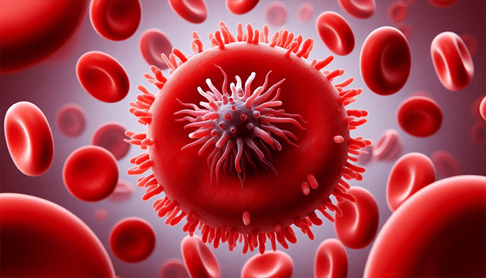
Morgellons Disease: What You Need to Know About Symptoms, Causes, and Treatment

Morgellons disease is a complex and often misunderstood condition that I’ve been treating for over 20 years. It’s marked by painful skin lesions that may contain colored fibers, and despite the severity of the symptoms, it’s frequently misdiagnosed. As a Seattle-based Lyme disease doctor, I’ve seen how infections like Lyme disease, Babesia, and Bartonella often contribute to Morgellons symptoms. This article aims to provide clarity on Morgellons disease, its potential causes, and the treatment options that have proven effective for my patients. For more information about my practice and approach to healing, visit my website.

Caption: Medical Minutes with Dr Marra, Episode 21 Morgellons Disease

What Is Morgellons Disease?

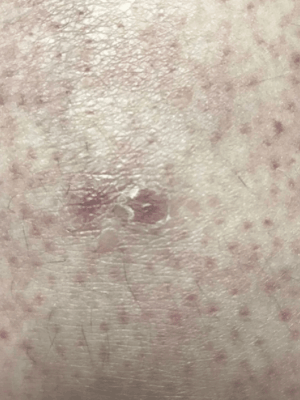
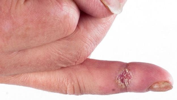
Morgellons disease is a debilitating skin condition characterized by painful, deep skin lesions that often contain visible colored fibers. These fibers are typically made up of keratin and collagen, though we still don’t fully understand why they appear in the skin. In my practice, I’ve seen more than 14,000 families affected by this condition, and it’s clear that Morgellons are real — despite the fact that traditional medicine has been slow to recognize it.

Key Symptoms of Morgellons Disease

  • Painful Skin Lesions: Often ulcerated and difficult to heal.
  • Colored Fibers: Visible within the lesions, usually composed of keratin and collagen.
  • Itching and Crawling Sensations: Many patients experience a sensation of movement under the skin, which adds to the distress.

Misdiagnosis: Why Morgellons Disease Is Often Not Taken Seriously

One of the biggest challenges with Morgellons disease is that it’s frequently misdiagnosed. Traditional doctors may incorrectly label it as “delusional parasitosis,” a psychiatric disorder, which is simply not the case. I’ve seen firsthand how this misunderstanding can make patients feel dismissed or ignored, which only adds to their suffering. Morgellons disease is a physical condition with identifiable symptoms, not a mental health issue.

What Causes Morgellons Disease?

Through my extensive experience, I’ve found that many patients with Morgellons disease test positive for several infections, including:

These infections, coupled with certain genetic factors, appear to be key contributors to the development of the painful skin lesions seen in Morgellons disease. However, the exact cause of Morgellons remains a mystery, and more research is needed to fully understand the underlying mechanisms.

Current Research on Morgellons Disease

There is a Morgellons Research Foundation, led by Cindy Casey, RN, who has been a tireless advocate for patients with this condition. Cindy herself suffers from Morgellons, which has given her a unique perspective on the disease. The foundation works to raise awareness and secure funding for research, although this has proven to be a challenging task.

If you’re looking for more information, I highly recommend watching the YouTube documentary Skin Deep, which sheds light on the struggles Morgellons patients face in convincing traditional doctors that this disease is real. Some progress is being made in research, particularly by scientists at Oklahoma State University, but there’s still much more to learn.

Diagnosing Morgellons Disease: What You Should Know

At present, there is no definitive skin test to confirm Morgellons disease. However, we can visualize the colored fibers under a high-powered microscope. In some cases, the fibers seem to move when a patient holds a starchy substance, such as a potato, in their hand. This suggests that the fibers could be linked to organisms that feed on sugar, though this idea is still not scientifically proven.

Effective Treatment for Morgellons Disease

In my 20+ years of treating Morgellons disease, I’ve found that a comprehensive, multi-pronged approach works best. Although treatment can be lengthy, often taking months or even years, my patients have seen significant improvement with a combination of therapies.

1. Antibiotics and Antiparasitics

The right combination of antibiotics and antiparasitic medications is often crucial in treating Morgellons disease. Here are a few that have worked for many of my patients:

  • Doxycycline (combined with Methylene Blue)
  • Ivermectin and Albenza
  • Dapsone Skin Cream

2. Topical Treatments for Skin Lesions

Topical creams can be effective for reducing inflammation and helping to heal the skin lesions. Anti-inflammatory dietary changes are also important in managing the condition.

3. Detoxification

Detoxing the body can support overall healing. I recommend strategies like staying hydrated, sweating, and using gentle detox methods to help rid the body of toxins, including mycotoxins.

4. Nutritional Support

Nutritional support is essential in helping the body heal from Morgellons disease. Key supplements that I recommend include:

  • Probiotics
  • Vitamin D
  • Curcumin
  • MSM
  • Fish Oil

These nutrients help support the immune system, reduce inflammation, and promote skin healing.

Early Diagnosis and Treatment from Dr. Susan Marra

I’ve seen firsthand how effective the right treatment can be in healing skin lesions and addressing the underlying infections that cause Morgellon disease. If you’re ready to take the next step toward healing, please don’t hesitate to contact us or call 206-299-2676. Together, we can create a personalized treatment plan to help you heal and regain your quality of life.

Disclaimer The information, including but not limited to, text, graphics, images and other material contained in these videos is for informational purposes only.  It is not intended to be a substitute for professional medical advice, diagnosis, or treatment. Always seek the advice of your physician before undertaking a new healthcare regimen, and never disregard professional medical advice or delay in seeking it because of something you have seen, read, or heard in these videos.


Lyme Disease in Dogs

What You Need to Know for Prevention and Treatment

As an avid pet lover, hearing “Lyme disease in dogs” may raise your concerns. Don’t worry, Dr. Marra is here to educate you on prevention, teach you to recognize the symptoms, and understand the treatment options available.

As most of you know, I am an avid dog lover and have had a dog in my office for most of my career, both for emotional support and mental health. Dogs can be excellent office companions on your health care journey. So you can imagine, I am very aware and concerned for dogs that get Lyme disease. I’m here to help.” 

Medical Minutes with Dr Marra; Episode 51: Lyme Disease in Dogs

What is Lyme Disease in Dogs?

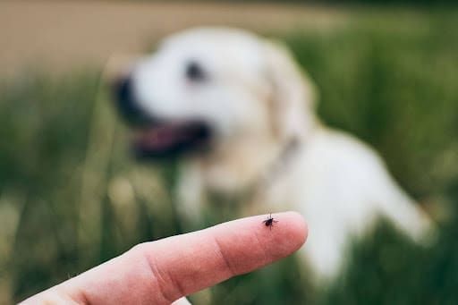
Lyme disease affects over 300,000 people annually in the United States and it CAN also affect dogs when they are bitten by an infected tick. The bacteria Borrelia burgdorferi, which causes Lyme disease, is transmitted through tick bites and can have significant effects on your dog’s health if not treated properly.

Symptoms of Lyme Disease in Dogs

Canine Lyme disease symptoms can vary, but they often include:

  • Generalized body pain
  • Limping
  • Swollen joints
  • Fever
  • Vomiting
  • Lethargy
  • Anorexia
  • Weight loss
  • Lameness

Unfortunately, it typically takes 2-5 months after the tick bite for dogs to show symptoms of Lyme disease, and by then, the bacterial infection has usually become systemic.

Diagnosing Lyme Disease in Dogs

Lyme disease can be diagnosed using blood tests for the bacteria antibodies (IgM and IgG) or a PCR test on blood or joint fluids. General blood work and a urinalysis can also help. The vet will look for signs of kidney damage, such as a loss of protein in the urine, which can indicate that the infection has affected the kidneys.

Treatment for Lyme Disease in Dogs

Treatment for Lyme disease in dogs is readily available and effective. Common medications include:

  • Doxycycline (the best choice)
  • Amoxicillin
  • Zithromax

Generally, your dog will need to stay on antibiotics for at least 4 weeks to completely eliminate the infection. This treatment is much more effective in dogs compared to humans, where treatment can be more challenging.

Lyme Disease Vaccine for Dogs

There is a vaccine available for dogs to help prevent Lyme disease. While it is safe and can reduce the risk of infection, it is NOT 100% effective. If you live in a Lyme-endemic area, discuss the pros and cons with your vet to determine whether this vaccine is appropriate for your dog. If you do not live in a Lyme-endemic area, your dog likely does not need this vaccine.

Tick Prevention for Dogs

Tick prevention is absolutely critical in Lyme-endemic areas. However, tick repellents for dogs can have side effects, so it’s important to discuss options with your vet. Some commonly used tick prevention products include:

  • Frontline
  • Advantix
  • Bravecto (topical)
  • Nexgard (chewable)
  • Simparica

Speak to your vet about which product would be best suited for your dog breed.

Be a Responsible Fur Parent: How to Protect Your Dog from Lyme Disease

Part of being an excellent fur parent is ensuring that your dog is protected from health dangers like Lyme disease, especially in endemic areas. It’s important to be aware of where ticks are common in your state/area, so you can take proactive steps to prevent Lyme disease.

Dogs are our dearest friends and allies, but they cannot protect themselves from Lyme disease. When they play outside, they are just being goofy dogs—but this is when they can be vulnerable to Lyme disease. To protect your canine companion, be sure to:

  • Discuss the prevalence of Lyme disease in your area with your vet
  • Use tick prevention medication
  • Stay vigilant in tick-prone areas

Your dog will love you for taking such great care of them!

FYI, NONE of my dogs have ever had Lyme disease because when I am in an endemic area, I generally use Frontline. I have never even found a tick on them. Thank goodness! I hope you take these precautions seriously to protect your beloved dog(s) from Lyme disease. Here’s to keeping both you and your furry friend healthy and happy!

About Dr. Susan Marra & Juju

Dr. Susan Marra is a world-leading Lyme Literate Naturopathic doctor specializing in the treatment of Lyme disease, vector-borne diseases, Neurodegenerative Diseases (Alzheimer’s, Parkinson’s disease), Post Covid, Long Covid, red light therapy, and environmental illness (mold).

Having trained with Dr. Richard Horowitz and Dr. Charles Ray Jones, Lyme disease experts on the East Coast, Dr. Marra is well qualified to manage and heal tick-borne disease. Take the first step toward a better quality of life with expert care tailored to your needs.

Juju (Juliette Pearl) is Dr. Marra’s cream french bulldog and the inspiration for understanding Lyme disease in dogs. Juju has wonderful healing energy and properties just because she is lovable and friendly and wants belly rubs all the time. If you join us in the office, you can find her treats at the front counter but please ONLY give her one.  We have to watch her weight because she is a true “bon bon” girl.

To learn more about Dr. Marra’s approach or to schedule a consultation, contact or call us to book an appointment. 


Disclaimer The information, including but not limited to, text, graphics, images and other material contained in these videos is for informational purposes only.  It is not intended to be a substitute for professional medical advice, diagnosis, or treatment. Always seek the advice of your physician before undertaking a new healthcare regimen, and never disregard professional medical advice or delay in seeking it because of something you have seen, read, or heard in these videos.

Oxalates and Kidney Stones

Oxalates are molecules found in certain foods such as spinach and cashews that are formed as the combination end product of several amino acids such as serine, glycine, and ascorbic acid, that can combine with calcium (a naturally occur ion in the blood also from food) in the blood to form calcium oxalate crystals.  Foods that contain more than 100g of oxalates per serving include (but this IS NOT an exhaustive list):

  • Beets
  • Rhubarb
  • Brussel Sprouts
  • Carrots
  • Spinach
  • Tofu
  • Rice Bran
  • Almonds
  • Snap Beans
  • Cashews
  • Soybeans
  • Quinoa
  • Blackberries
  • Blueberries
  • Figs
  • Plum
  • Raspberry
  • Tangerine
  • Sweet Potatoes
  • Eggplant
  • Kale

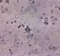
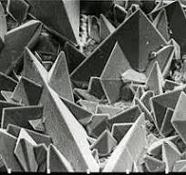
These oxalate crystals are needle shaped crystals called raphides.  They are readily absorbed after ingestion, cannot be metabolized by mammals, and are largely eliminated through renal excretion.  Calcium oxalate crystals get filtered by the glomeruli in the kidney unless they are large, and then they become “stuck” in the nephrons and form kidney stones, especially if you have the genetic propensity.  Oxalates are toxic to the renal epithelial cells and can cause membrane injury.  The common upper limit of normal for oxalate excretion is 45mg/day (0.5mmol/day).  Chronic vitamin C oral use can also cause hyperoxaluria and kidney stones. 

Kidney stones are EXTREMELY painful, and the symptoms associated with them are as following:

SEVERE and sudden onset pain on the abdomen sides or low back

  • Nausea
  • Vomiting
  • Fever
  • Chills
  • Blood in the urine
  • Cloudy urine

Small kidney stones can be passed in the urine, but larger ones greater than 9mm must be treated with lithotripsy (a procedure where a skilled physician blasts the  stones into pieces for excretion, or surgery).  The pain associated with kidney stones is very significant and usually requires pain management by the ER.

To check for oxalate crystals in the urine, you can have your doctor order a 24 hour urine oxalate test through Labcorp or Quest, and this will reveal urinary levels and the presence of toxicity.

Treatment for calcium oxalate crystals is largely dietary.  You’ll need to go on a low oxalate diet, and read the book “Toxic Superfoods” by Sally Norton, MPH.  She has some excellent information on how to change your diet if you are prone to kidney stones.

I was plagued by kidney stones 10 years ago from over eating spinach and cashews and the urologist NEVER told me to change my diet.  After researching the subject, I found that you can lead a very normal life IF you curb your diet from high oxalate foods, and drink at least 64oz of water with electrolytes every day.

Professional Health Products has two supplements, Oxalate Scavenger, and Oxalate Balancer that contain nutrients such as: Calcium citrate, Magnesium oratate, vitamin B6 (P5P) or Calcium, Magnesium, and Zinc citrate with P5P which will bind the oxalate crystals.

Kidney stones are no fun and you can avoid them simply by being mindful with your diet.  Find a list with high oxalate containing foods, and remove the top 10-15 of them completely from your diet and that should do the trick.  Make sure you drink plenty of fresh water to cleanse the kidneys daily and you should be just fine.

Of note, it is my observation that patients with Bartonella are much more likely to develop kidney stones although to date, this is an anecdotal observation.  I happen to be one of these patients.

Understanding Hypermobile Ehlers-Danlos Syndrome (hEDS)

Joint Pain HEDS

Understanding Hypermobile Ehlers-Danlos Syndrome (hEDS)


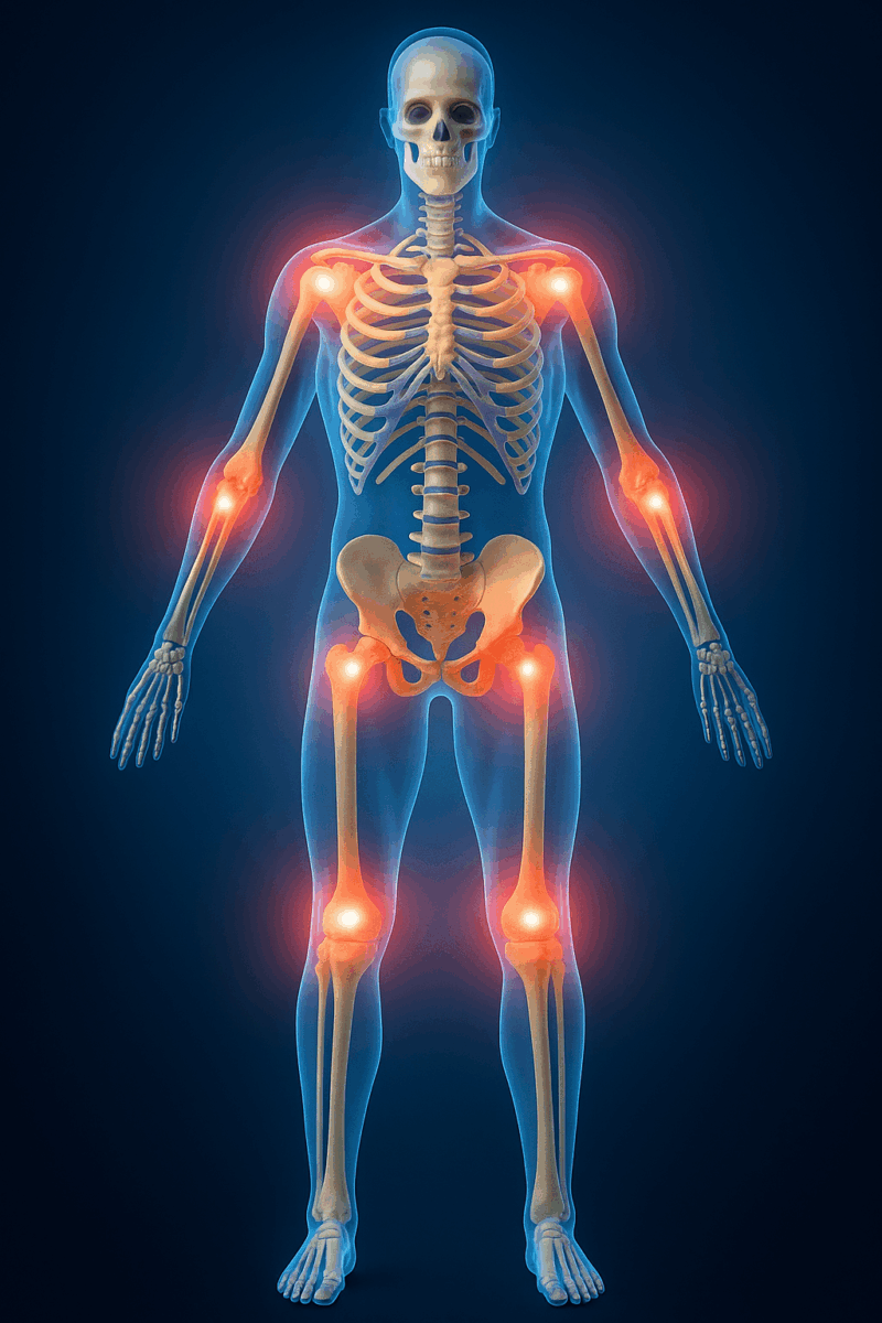
Hypermobile Ehlers-Danlos Syndrome (hEDS) is a genetic disorder that affects the body’s connective tissues, specifically collagen. Collagen is a crucial protein that provides structure and elasticity to skin, joints, blood vessels, and organs. In people with hEDS, a defect in collagen production results in weakened connective tissues, leading to a variety of symptoms, including hypermobile joints, fragile skin, joint pain, and easy bruising.

Dr. Marra is currently diagnosed with hEDS, which gives me a deep understanding and comprehension of its impacts on day-to-day life. It also enables the ability to diagnose it more accurately. Watch the Medical Minutes video below for a full breakdown of hEDS.

What is hEDS?

hEDS is one of 13 types of Ehlers-Danlos Syndrome (EDS), a group of genetic disorders that involve abnormalities in collagen. Collagen is a key structural protein, and when it’s defective, it can affect the stability of the body’s connective tissues. In hEDS, individuals often experience hypermobile joints, which are joints that stretch beyond the normal range of motion and skin hyperelasticity, which means the skin is unusually stretchable and fragile. These abnormalities can lead to frequent joint dislocations, pain, bruising, and delayed wound healing due to abnormal collagen production.

Symptoms of hEDS

The symptoms of hEDS can vary widely but typically include:

  • Joint Hypermobility: An increased range of motion in the joints, which may cause pain, dislocations, and instability.

  • Chronic Pain: Due to overstretched ligaments and muscles, individuals often experience joint pain and frequent injuries.
  • Skin Fragility: The skin may be unusually elastic and prone to bruising and tearing.
  • Fatigue: Chronic fatigue is common, likely due to the strain on muscles and joints.
  • Abnormal Scarring: Due to defective collagen, scars from injuries or surgeries may heal poorly and be more prominent.
  • Frequent Bruising: Bruises may appear even with minimal trauma, as the skin’s fragility increases the likelihood of bruising.

Histamine Intolerance and hEDS

An interesting aspect of hEDS involves histamine intolerance, which can make symptoms worse. Histamine is a chemical involved in immune responses and inflammation. In people with hEDS who also have allergies or excessive histamine production, histamine can degrade collagen, worsening symptoms like joint pain and skin irritation. This can lead to flare-ups where pain, swelling, and fatigue are exacerbated. Managing histamine levels through diet, antihistamines, and avoiding allergens can help alleviate these flare-ups and improve overall symptom control.

Treatment and Management of Hypermobile EDS

Currently, there is no cure for hEDS, but the condition can be managed with supportive therapies. Treatment focuses on relieving pain, improving joint stability, and managing other symptoms.Early diagnosis and a tailored care plan are key to managing this condition effectively. Common treatment options include:

  • Physical Therapy (PT): Strengthening muscles around hypermobile joints helps stabilize them and reduce pain.
  • Strain-Counterstrain Therapy: This specialized technique helps alleviate muscle tension and reduce pain by positioning the body to relieve strain.
  • Pain Management: NSAIDs and other medications can be used to manage pain and inflammation.
  • Joint Protection: Splints, braces, and other devices can help protect hypermobile joints from injury.

Get Expert Care for hEDS: Schedule a Consultation with Dr. Susan Marra

If you or someone you know is struggling with hEDS or related connective tissue disorders, it’s important to seek specialized care. Dr. Susan Marra, an expert Naturopathic Doctor and Master of Science Psychology, offers comprehensive treatment options to help manage the symptoms of hEDS and improve your overall well-being. Dr. Marra combines the latest research with personalized care plans to address the unique needs of each patient. To learn more about Dr. Marra’s approach or to schedule a consultation, contact or call us to book an appointment. Take the first step toward a better quality of life with expert care tailored to your needs.

Book an Appointment

About Dr Marra

About Dr. Susan Marra

Dr. Susan Marra is a world-leading Lyme Literate Naturopathic doctor specializing in the treatment of vector-borne diseases (Lyme, Bartonella, Babesia, Relapsing Fever, Ehrlichia, Anaplasma, Tularemia, Q Fever, Rocky Mountain Spotted Fever, Rickettsia Species), Neurodegenerative Diseases (Alzheimer’s, Parkinson’s disease), Post Covid, Long Covid, anti-aging, and environmental illness (mold).

Having trained with Dr. Richard Horowitz and Dr. Charles Ray Jones, Lyme disease experts on the East Coast, Dr. Marra is well qualified to manage and heal tick-borne disease.

Disclaimer The information, including but not limited to, text, graphics, images and other material contained in these videos is for informational purposes only. It is not intended to be a substitute for professional medical advice, diagnosis, or treatment. Always seek the advice of your physician before undertaking a new healthcare regimen, and never disregard professional medical advice or delay in seeking it because of something you have seen, read, or heard in these videos.

Continue reading

Photobiomodulation (PBM) Information Sheet

Photobiomodulation (PBM) Information Sheet

September, 2024

Molecular Mechanisms of Alzheimer disease:

  • decreased cognitive functioning
  • decreased capacity to learn new information
  • decreased memory capacity
  • decreased ability for decision making
  • decreased language abilities
  • decreased motor activities
  • decreased mitochondrial functioning due to decreased enzyme cytochrome c oxidase (CCO) activity
  • abnormal deposition of Beta-Amyloid protein in the brain
  • imbalance of mitochondrial fusion and fission proteins
  • increased neuronal inflammation by microglial and astrocyte cell activity
  • induction of heat shock proteins
  • induction of cyclooxygenase pathway
  • NF Kappa beta stimulated neuritis
  • increased oxidative stress from increased reactivity oxygen species (ROS)
  • decreased ATP production by the mitochondria
  • increased expression of neurofibrillary tangles

Benefits of Photobiomodulation ( PBM) in Alzheimer disease:

  • transcranial application of:
  • red light (600-770nm)
  • near infrared light (760-1200nm)
  • removes deposition of Beta-Amyloid protein in the brain
  • induces changes in the genetic expression BACE1 and Cathepsin B enzymes
  • inactivates JNK3 gene that lead to decreased endocytosis resulting in less beta-amyloid protein deposition
  • 1070nm NIR depletes beta-amyloid deposition
  • improves cognitive functions such as memory and learning
  • promotes vasodilation by increasing cerebral blood flow
  • this removes metabolic waste
  • helps remove beta-amyloid breakdown debris
  • disaffiliates the nitric oxide (NO) inhibitor and thereby increasing the availability of cytochrome C oxidase (CCO) enzyme in maintaining mitochondrial membrane function and potential
  • increases oxygen consumption
  • increases glucose metabolism
  • increases ATP biosynthesis by the mitochondria
  • regulates mitochondrial homeostasis
  • allows for recovery of damaged human neuronal progenitor cells by maintaining mitochondrial homeostasis
  • enhances cytochrome C oxidase activity (CCO)
  • increases the efficiency of ATP production by the mitochondria
  • improves the flow of electrons through the electron transport chain
  • increases mitochondrial membrane potential
  • specifically stimulates the activity of complex VI in the mitochondria
  • promotes new mitochondria generation
  • lowers inflammatory response
  • decreases neuronal degradation
  • shifts the microglial phenotype from M1 (proinflammatory) to M2 (antiinflammatory)
  • decreases Beta-Amyloid production
  • decreases the expression of inflammatory markers in Alzheimer disease
  • modulates proinflammatory cytokines such as tumor necrosis factor alpha (TNF alpha)
  • near infrared (NIR) light decreases microglial toxicity
  • near infrared (NIR) light promotes neuron survival
  • decreases oxidative stress from reactive oxygen species (ROS), toxins, heavy metal, etc…
  • increases antioxidant enzyme capacity
  • increase the positive aspects of reactive oxygen species (ROS) by provoking cytoprotective, antioxidant, and anti-necrotic effects of neurons
  • increases cerebral blood flow through nitric oxide (NO) mediated vasodilation
  • decreases depression by altering glutamatergic dysfunction by regulating GLT-1 mRNA protein
  • enhances tissue repair
  • utilizes neuroprotective properties
  • stimulates neurogenesis (formation of new neurons)
  • promotes synaptogenesis (formation of new synapses)
  • promotes neural repair and functional recovery
  • influences neuronal activity by altering ion channel functioning and neurotransmitter release
  • has a positive effect on the rest-activity rhythm of Alzheimer disease

Benefits of Photobiomodulation (PBM) in Traumatic Brain Injury (TBI):

  • improves cognitive function
  • decreases post concussive symptoms
  • accelerate recovery in patients with mild-moderate traumatic brain injury (TBI)
  • neuroprotective effects
  • anti-inflammatory effects

Benefits of Photobiomodulation (PBM) in Stroke:

  • enhances neurogenesis (growth of new neurons)
  • decreases brain damage
  • improves functional recovery
  • improves motor function
  • enhances rehab outcomes

Benefits of Photobiomodulation (PBM) in Parkinson disease:

  • neuroprotective from continued loss of dopaminergic neurons in the substantia nigra part of the brain
  • may lower the accumulation of alpha synuclein within neurons called Lwey Bodies
  • may heal vascular damage from proinflammatory cytokines
  • may heal blood brain barrier leakage from proinflammatory cytokines
  • may heal the gut microbiome
  • increases mitochondrial function by increasing ATP production
  • lowers microglial activation from inflammation
  • may restore gut motility and vagus nerve activity
  • may reduce alpha synuclein accumulation in the gut
  • reduces inflammation by lowering proinflammatory cytokine levels such as TNF alpha, interleukin 1 (IL-1), interleukin 6 (IL-6)
  • decreases neuronal damage
  • slows disease progression
  • improves cognitive function
  • improves motor and mobility performance (i.e., walking, gait, tremors, writing, fine motor control)
  • may improve sense of smell
  • may improve mood
  • may improve social interactions
  • influences the gut microbiome to reduce alpha synuclein accumulation in the gut
  • may improve glymphatics which helps remove toxins from the brain
  • may improve confidence
  • may improve balance
  • improves angiogenesis (new blood vessel formation) 
  • improves collateral and capillary revascularization
  • improves blood flow
  • improves blood filling in the cerebral hemispheres
  • decreases gliosis (scarring in the brain)
  • induces neurogenesis (new neuron formation)
  • induces regenerative cerebral changes

Benefits of Photobiomodulation (PBM) for Pain:

  • analgesic effect on pain
  • decreases inflammation
  • increases tissue repair
  • decreases neuropathic pain by decreasing inflammation

Benefits of Photobiomodulation (PBM) for Depression:

  • influences brain activity of glutamatergic neurons
  • promotes neuroplasticity
  • has mood modulating effects
  • used in conjunction with pharmaceuticals and NOT in place of meds

Excerpted from: 

“Unleashing light’s healing power: an overview of photobiomodulation for Alzheimer treatment”. Ramanishankar, A., Singh, A., Begum, R., Jayasankar, N., Prajapati, B., Nirenjen, S.  Future Science OA, 10:1/ fsoa-2023-0155, 2024.

“Parkinson’s Disease and Photobiomodulation: Potential for Treatment.”  Bicknell, B., Liebert, A., Herkes, G.  Journal of Personalized Medicine, 2024, 14, 112, pages 1-32.

“Photobiomodulation for the Brain.  Photobiomodulation Therapy in Neurology and Neuropsychiatry”  Salehpour, F, Sadigh-Eteghad, S., Mamoudi, J., Kamari, F., Cassano, P., Hamblin, M. Springer, 2023.

“Photobiomodulation in the Brain.  Low Level Laser Light Therapy in Neurology and Neuroscience.”  Edited by: Hamblin, M., Huang, Y.Y.  Elsevier Academic Press, 2019.

Dr. Bob Bransfield – Neuropsychiatric Manifestations of Tick Borne Infections

CRYPTO-INFECTIONS CONFERENCE 2023

LYME DISEASE & OTHER HIDDEN INFECTIONS: MICROBIAL PERSISTENCE

Robert C. Bransfield, MD, DLFAPA

The Third international ‘CryptoConference’ was held in Dublin Ireland June 17th to 18th 2023. ‘Crypto’ refers to occult infections which are hard to diagnose and hard to treat. The theme of the third conference is ‘microbial persistence’, which is the way that these occult infections evade the immune system and evade routine diagnostic techniques. We invited a range of clinicians and basic scientists from the USA, Canada, and throughout the European Union, to spend two days in Dublin. The topics to be addressed include Borrelia, Bartonella, Babesia, and a range of other microbial pathogens. Lectures provided will appeal to primary care doctors (general practitioners) on the subject of Lyme and Long COVID, as well as more basic science lectures that will appeal to bench researchers as well as clinicians.

The Importance of Probiotics

The importance of probiotics while undergoing Lyme Disease treatment cannot be underestimated. The human microbiome is composed of trillions of different bacteria that live harmoniously and perform various necessary physiological functions, especially in the intestines. These bacteria have specific nutritional needs just as we do to sustain life. Additionally, endigenous bacteria generally do not cause disease, unless the bio-terrain is altered by other infectious organisms, antibiotics, or poor diet. In this case, your “garden” becomes a “garden of weeds.”

While antibiotics are necessary to eradicate Lyme Disease and co-infections in most patients, great care should be taken to maintain a healthy microbiome while undergoing treatment. A plant-based diet low in carbohydrates and plenty of water keeps inflammation at a minimum so that the antibiotics can work optimally to kill pathogenic organisms.

But make no mistake…this is risky business because antibiotics not only kill the bad organisms, they also kill the good ones. Therefore, high-dose, diverse, probiotics should be taken alongside antibiotic therapy without question. Some of these can include:

Lactobacillus acidophilus

Lactobacillus brevis

Lactobacillus plantarum

Bifidobacterium lactis

Bifidobacterium longum

Saccharomyces boulardii

Additionally, pre-biotics in the form of FOS (fructooligosaccharides) may be necessary to balance the gut flora.

As you age, your gut microbiome becomes increasingly less able to optimally function. This gets exacerbated when you have multiple infections affecting the mucos.

Methylene Blue

Methylene Blue is an old drug (circa 1876) used to treat methemoglobinemia and cyanide poisoning.  It is a thiazide dye and turns your urine blue, but generally is very well tolerated. Some patients like to use red spectrum light with methylene blue, especially for treating plaque psoriasis.

It works by converting ferric iron in hemoglobin to ferrous iron.  It also has anti-inflammatory properties and is currently under research investigation for the treatment of persistent Bartonella with promising results.

The only caveat is that you must be tested for G6PD (glucose-6-phosphate-dehydrogenase) deficiency to ensure that enzyme levels are normal, prior to beginning treatment.
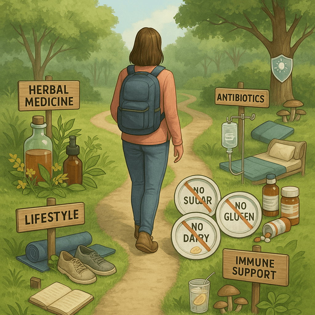
Sleep and Insomnia

Sleep is critical for healing, particularly deep sleep. During sleep, the body alternates between REM sleep and non-REM sleep.  During REM or deep sleep, you dream, and dreaming is thought to be the way that the mind processes information. During sleep, most of the body’s systems are in an anabolic state, helping to restore the immune, nervous, muscular, and skeletal systems.  Sleep is also the time that the brain detoxes through its glymphatic system.

Insomnia is a common symptom of tick-borne disease and is characterized by an inability to fall asleep, stay asleep, and wake refreshed.  This has to be treated right away so that you can get 8-10 hours of restful sleep a night.  

There are many supplements that can help with sleep 30 minutes before bed includes:

  • Melatonin (1-4 mg)
  • Phosphatidylserine (100-400 mg)
  • L-Theanine (750 mg)
  • DMG (Dimethylglycine) (100 mg)
  • GABA (gamma-Aminobutyric acid) (1000 mg)
  • Valerian (100 mg)
  • Progesterone (25 mg – 100 mg)
  • 5-HTP (50 mg – 100 mg)

There are also several peptides that are sleep aids including:

  • Epitalon
  • DSIP (Delta sleep-inducing peptide)

Lastly, medications that can help with sleep include:

  • Trazodone
  • Quetiapine
  • Diphenhydramine
  • Doxepin
  • Hydroxyzine
stages of sleep

Detoxification

Detoxification is a natural property of the human body and includes urination, defecation, sweating, and breathing. Food and liquids that we eat and drink respectively contain many components for which the body cannot use and are therefore eliminated as waste. This is a homeostatic component of our physiology that allows “balance” to be maintained in the body.

The liver, kidneys, colon, and lungs are the organs responsible for carrying out detox activity in the body. However, the liver bears most of this burden in filtering blood. Nature has designed four elaborate detoxification pathways that include: sulfation, glucuronidation, acetylation, and glycation. These pathways must operate with ease in order for the liver to filter contaminants in the blood such as spirochetal excrement, dead spirochetes, and known toxins such as Bb Tox 1, ammonia, and Quinolinic acid. If these toxins are not removed from the body, they accumulate causing widespread inflammation and tissue damage which augments illness and may contribute significantly to chronic infectious illness.

detoxification remedies

Healing Affirmations

Healing Affirmations are an incredibly powerful tool in the healing journey. One of the things that are so disheartening about chronic illness is that your spirit can become sick and depressed as well.  This complicates the healing journey substantially.  One way to keep your spirits high while you heal from chronic illness is to do daily affirmations that actually help you heal.  Below are affirmations that you can do or write in your journal to keep your spirits high:

  • I know that I am healing every day
  • My cells feel better and better every day
  • My infections are getting treated every day
  • I know that my body will be well very soon
  • I can feel my body healing every day
  • I am not sick. I am just having a bad day
  • I am just resting to allow my body to heal, which will happen quickly
  • I am so grateful for my herbs and medications that help me heal
  • I am so grateful that I can move, eat, and spend time with my family
  • I am so grateful that I still have a good attitude and goals that I want to accomplish

There are many books online with healing affirmations that you can purchase.  Do this daily, and it will make your healing time shorter. Psychoneuroimmunology is a significant factor in your return to health ad you are in control of that.

Another great way to start your day is with a gratitude journal. Every morning, write down 10 things that you are most grateful for and make them different every day. By doing this, you begin to change your frequency to a higher vibration which is always a better option when you are trying to heal from chronic illness.

healing affirmations

Cannabis for Lyme Symptoms

cannabis leaves

If you are having trouble managing the symptoms of Lyme disease or other co-infections, medical cannabis may be a treatment option for you. The anti-inflammatory properties of medical cannabis have been linked to providing potential relief for some of the symptoms of Lyme disease, as well as numerous mental benefits including some relief from anxiety and depression. The pain-relieving properties of medical marijuana make it a viable treatment for Lyme disease. Cannabis is a naturally occurring herb known for its relaxing and neurologically calming properties and has recently become available in Washington State for medicinal use. Cannabis contains Cannabidiol (CBD) which is the major non-psychoactive constituent of Cannabis sativa, and Delta 9 – tetrahydrocannabinol (THC), which does affect mental acuity.

Cannabinoids promote neurogenesis and have neurological protective effects as well as pain-reducing effects on the body. Therefore, Cannabinoids may be very useful in the treatment of chronic tick-borne illness. The key is to use an extract that is high in CBD but low in THC, so that anti-inflammatory properties are maximized, and mental “dulling” properties are minimized.

If you would like to know more about Cannabinoids in the treatment of chronic tick-borne illness, please contact Dr. Marra. Not everyone is a candidate to use this herb, and so a detailed history and physical will be required to assess your eligibility for this treatment.
Anti-Inflammatory Diet

Inflammation is the body’s normal response to infections, diseases, injuries, and anything it considers harmful.  It is important to lower inflammation during treatment because this will help fight against toxic compounds, pathogens, and damaged cells build up in organ tissues. Certain foods are known to cause inflammation in the body. One of the fastest ways to decrease inflammation is to adopt an anti-inflammatory diet.

Fruits and vegetables are particularly important in the defense against inflammation. Vegetables in the allium family include garlic, scallions, onions, Chinese chives, and leeks.

Cruciferous vegetables include broccoli, cabbage, cauliflower, mustard greens, and brussel sprouts.

Phytonutrients are found in all berries and colorful fruits. They are naturally sweet and a great alternative to sugar.

Beans are a great source of protein and contain important minerals such as magnesium, potassium, zinc, and iron. These include black beans, red kidney beans, navy beans, garbanzo (chickpeas) beans, and pinto beans. You should eat these at least twice a week.

Here are anti-inflammatory foods to include in your diet: Blueberries, Blackberries, Cherries, Dark Grapes, Kale, Spinach, Cauliflower, Brussels Sprouts, mustard greens, cabbage, scallions, leeks, broccoli, beans, lentils, onions, garlic, green tea, avocado, coconut, olives, walnuts, pistachios, pine nuts, almonds, turmeric, ginger, cinnamon, dark chocolate, fish, spices

anti-inflammatory foods

Alkalizing Foods

The alkaline diet is based on the idea that replacing acid-forming foods with alkalizing foods can improve your health. The body fluids of healthy individuals are naturally alkaline meaning they have a high pH, whereas the body fluids of the chronically ill tend to be more acidic with a low pH. Most degenerative diseases attributed to aging, cancer, osteoporosis, and heart disease have all been scientifically linked to calcium and other mineral deficiencies that result in the body fluids becoming acidic. Acidosis is a common factor in over 150 degenerative diseases. Adopting more alkalizing foods into your diet can have positive impacts on your overall health and help you to along the detoxification process.

Put simply, pH is a measurement of how acidic or alkaline something is.

The pH value ranges from 0–14:

  • Acidic: 0.0–6.9
  • Neutral: 7.0
  • Alkaline (or basic): 7.1–14.0

Incorporate Alkaline foods including:

  • Fruit
  • Unsweetened fruit juices
  • Raisins
  • Black currants
  • Vegetables (especially spinach)
  • Potatoes
  • Wine
  • Mineral soda water
  • Soy food
  • Legumes
  • Seeds
  • Nuts

Neutral foods: natural fats, starches, and sugars

Finally, people interested in trying an alkaline diet should avoid high-acid foods. These include:

  • dairy products such as cheese and milk
  • processed foods
  • fish
  • coffee
  • alcohol
  • soda
  • salt
bowl of greens

Oxidative Stress and Mycotoxins

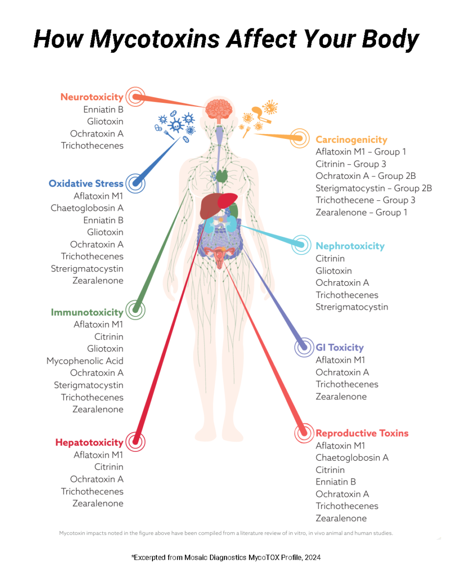
Mycotoxins can cause oxidative stress. Oxidative stress is an intracellular imbalance that occurs in chronic disease where reactive oxygen species are not readily removed and detoxified into more inert compounds in the body. This results in cellular dysfunction leading to disease. The production of free radicals and peroxides are chemicals that cause intracellular organelle damage and prevent normal cellular function. Mycotoxin exposure significantly increases oxidative stress and therefore reduces glutathione. When glutathione is low, disease ensues.

However, glutathione can be replenished by an oral paste or liquid very effectively. When glutathione levels return to normal, proper cellular function ensues and often disease-causing symptoms diminish substantially. For more details please ask Dr. Marra for more details on how mycotoxins can cause oxidative stress in your next appointment.

Mycotoxins and Mold Exposure

Mycotoxins and mold are metabolites of fungal organisms that are toxic to the human body and exposure can be present in both food and air. Two food-borne mycotoxins are Aflatoxin and Ochratoxin. Aflatoxin is produced by many species of Aspergillus fungi and can be found in soil, peanut butter, olive oil, and some cosmetics. Ochratoxin is also produced by Aspergillus species and can be found in cereal, coffee, and wine. It too has disease-causing effects on the body and has a half-life of 35 days.

In September 2012, Dr. Dennis Hooper, director of Realtime Laboratories in Carrollton, Texas, presented information on the new technology available to investigate mycotoxins and mold exposure in patients. He has developed a urine PCR test for identifying mycotoxins, which are known causative agents in many chronic diseases and probably complicate healing from Lyme disease.

Additionally, Trichothecenes are airborne mycotoxins produced by several molds but most notably, Stachybotrys, otherwise known as “black mold.” Stachybotrys is a mold that arises from damp conditions and water leakage in homes. These toxins can produce significant irritation to the mucosal lining of the lungs, GI tract, bladder, and brain causing chronic illness. Patients exposed to Trichothecenes often report having severe headaches and brain fog which lessen when they change to a mold-free environment.

However, mycotoxins can get trapped in the body by adhering to fat cells or entering cells (especially adipocytes) causing oxidative stress. Oxidative stress for prolonged periods of time depletes intracellular glutathione and interferes with the cell’s ability to self-repair.

Herxheimer Reaction (HERX)

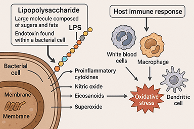
The Jarisch-Herxheimer reaction was originally noted in 1895 by Dr. Jarisch who worked with Syphyllis (a spirochete) patients.  In the 21st century, we observe Herxheimer reactions in tick-borne illnesses such as Lyme Disease, also caused by a spirochete.  A Herxheimer reaction (Herx) typically occurs hours to days after antimicrobial administration and is thought to be the result of endotoxins released into the bloodstream by dying spirochetes.  The body’s immune system then responds to the endotoxins and the concurrent release of cytokines (i.e., tumor necrosis factor, interleukin 6, and interleukin 8) which are chemical messenger molecules of the immune system producing exaggerated symptoms lasting 3 days up to several weeks.

Dr. Marra doses antibiotics specifically for the individual patient so that the Herxheimer reaction on a scale of 1-10, is about a 5.  This is her own technique, and she feels that severe Herxheimer reactions over prolonged periods of time may cause tissue damage from the excessive release of endotoxins and cytokines.  Tissues become friable as the result of chronic inflammation, not unlike what happens when a sponge is repeatedly used and the surface area becomes frayed.  Dr. Marra is not convinced that this kind of tissue damage is reversible which is why she chooses to take the route of treatment that she does.  However, many other physicians choose to treat more aggressively and seem to have good results. This is simply her own treatment preference.

The general description of a Herxheimer reaction is a temporary increase in symptoms but may also include the development of completely new symptoms.  More specifically, the most common complaints of a Herx reaction are increased joint and muscle pain, headaches, chills, night sweats, fever (usually low grade), extreme fatigue, and brain fog or lack of mental clarity, drop in blood pressure, hives, or a rash of some sort.  Worth noting here is that a rash or hives can sometimes be mistaken for an allergic reaction.  It is up to the treating physician to determine this, but with close, experienced observation and the use of Benadryl, Alka Seltzer Gold, Quercitin, warm lemon water and vitamin C (see Herx management), unnecessary premature cessation of antibiotics may be prevented.  In more severe cases of Herxheimer reactions, a reduction in the antibiotic dosage or cessation of the antimicrobials may be required.

The timing of a Herx reaction is very individualistic and can occur within days to weeks after the onset of antibiotic treatment.  In some patients, a Herxheimer reaction occurs only once or twice, or not at all throughout treatment.  It is clear however, that as you move through the treatment and the spirochetal load becomes decreased, the Herx reactions become less and less severe.  Herxheimer reactions can also occur in cycles (usually 4 week cycles) and may be affected by the lunar cycle.  This is anecdotal but it is an interesting observation in some people.

About 70-80% of people undergoing Lyme Disease treatment with antibiotics experience some sort of Herxheimer reaction.  It is thought by a number of physicians and scientists that these people have difficulty in detoxification processes, and that perhaps there may be a defect in one of the detox pathways in the liver.  This may be genetic, environmental or a combination of both.  But without question, maintaining an alkaline diet and an  “alkaline bioterrain” helps lessen the effects of a Herx reaction.  The 20% of patients who do not experience a Herx reaction should not feel like they are getting nowhere by taking antibiotics.  Discussion of this issue should be left to the patient and their treating physician.

Check out our page about alkalizing foods.

Herxheimer Reaction

The Herxheimer reaction (HERX) was originally noted in 1895 by Dr. Jarisch who worked with Syphyllis (a spirochete) patients.  In the 21st century, we observe Herxheimer reactions in tick-borne illnesses such as Lyme Disease, also caused by a spirochete.  A Herxheimer reaction (Herx) typically occurs hours to days after antimicrobial administration and is thought to be the result of endotoxins released into the bloodstream by dying spirochetes.  The body’s immune system then responds to the endotoxins and the concurrent release of cytokines (i.e., tumor necrosis factor, interleukin 6, and interleukin 8) which are chemical messenger molecules of the immune system producing exaggerated symptoms lasting 3 days up to several weeks.

Dr. Marra doses antibiotics specifically for the individual patient so that the Herxheimer reaction on a scale of 1-10, is about a 5.  This is her own technique, and she feels that severe Herxheimer reactions over prolonged periods of time may cause tissue damage from the excessive release of endotoxins and cytokines.  Tissues become friable as a result of chronic inflammation, not unlike what happens when a sponge is repeatedly used and the surface area becomes frayed.  Dr. Marra is not convinced that this kind of tissue damage is reversible which is why she chooses to take the route of treatment that she does.  However, many other physicians choose to treat more aggressively and seem to have good results. This is simply her own treatment preference.

The general description of a Herxheimer reaction is a temporary increase in symptoms but may also include the development of completely new symptoms.  More specifically, the most common complaints of a Herx reaction are increased joint and muscle pain, headaches, chills, night sweats, fever (usually low grade), extreme fatigue, brain fog or lack of mental clarity, drop in blood pressure, hives, or a rash of some sort.  Worth noting here is that a rash or hives can sometimes be mistaken for an allergic reaction.  It is up to the treating physician to determine this, but with close, experienced observation and the use of Benadryl, Alka Seltzer Gold, Quercitin, warm lemon water, and vitamin C (see Herx management), unnecessary premature cessation of antibiotics may be prevented.  In more severe cases of Herxheimer reactions, a reduction in the antibiotic dosage or cessation of the antimicrobials may be required.

The timing of a Herx reaction is very individualistic and can occur within days to weeks after the onset of antibiotic treatment.  In some patients, a Herxheimer reaction occurs only once or twice, or not at all throughout treatment.  It is clear, however, that as you move through the treatment and the spirochetal load becomes decreased, the Herx reactions become less and less severe.  Herxheimer reactions can also occur in cycles (usually 4-week cycles) and may be affected by the lunar cycle.  This is anecdotal but it is an interesting observation in some people.
About 70-80% of people undergoing Lyme Disease treatment with antibiotics experience some sort of Herxheimer reaction.  It is thought by a number of physicians and scientists that these people have difficulty in detoxification processes and that perhaps there may be a defect in one of the detox pathways in the liver.  This may be genetic, environmental, or a combination of both.  But without question, maintaining an alkaline diet and an  “alkaline bio terrain” helps lessen the effects of a Herx reaction.  The 20% of patients who do not experience a Herx reaction should not feel like they are getting nowhere by taking antibiotics.  Discussion of this issue should be left to the patient and their treating physician.

Check out our page about alkalizing foods.

Dyslipidemia and Lyme Disease

Dyslipidemia is abnormal lipid metabolism. Patients who have had or have Lyme Disease often have significant abnormalities in their lipid metabolism.  This results from chronic inflammation and prolonged activation of the proinflammatory cytokines such as IL-1, IL-6, and tumor necrosis factor-alpha (TNF alpha).  Elevated cholesterol, triglycerides, VLDL, and LDL are commonly seen, while HDL (the good cholesterol) is decreased.  Dr. Jones and I believe that these elevations in cholesterol are not only related to diet and genetic predisposition but may be a compensatory response of the body in an attempt to repair cell membranes (that are composed of lipids) that have been damaged by infection. The piercing of the cell membrane by organisms such as Borrelia, Bartonella, and Mycoplasma on their way to the intracellular compartment where they prefer to reside, can render changes in lipid metabolism and increase lipid peroxidation.  Lipid peroxidation is a biochemical reaction resulting from damaged lipid-containing cells that increases the concentration of free radicals in the body and contributes to a state of overall oxidative stress.  Oxidative stress affects not only cells but the vascular endothelium which can lead to increased stroke and myocardial infarction.  The presence of biofilm created by spirochetes and other organisms also contributes to plaque formation in the arteries, which affects blood flow to peripheral tissues and the overall health of the cardiovascular system.

Optimizing lipid metabolism may require diet and lifestyle modifications as well as nutritional intervention.  Dr. Marra’s extensive knowledge and experience in Lyme Disease and Functional Medicine will help you to regain optimal health in a reasonable amount of time.

For more detailed information about the glycemic index of foods (how much sugar they contain), please visit www.Mendosa.com and go to the food tab. You can search through the various options. This website is a phenomenal resource for everyone who wants to change their diet and needs help understanding what foods they can and can’t eat to improve their metabolic condition.

Gestational Lyme Disease

Unfortunately, there is great debate within the medical community regarding the existence and prevalence of the transmission of Lyme disease, co-infections, and parasites to the unborn fetus.

The medical community already recognizes that certain infections cross the placental barrier and they are known as “TORCH Syndrome” infections which include: Toxoplasmosis, Other (i.e., Syphilis), Rubella, Cytomegalovirus, and Herpes Simplex Virus. These infections become of great concern to the physician when unexplained birth defects occur in newborn babies. Antibody titer blood tests for “TORCH” infections in these babies yield insights into the cause of the birth defects, and this phenomenon is well established in the infectious disease medical community.

Operating purely from a perspective of common sense, if Syphilis crosses the placental barrier and can cause birth defects, why wouldn’t Borrelia, since they are both spirochetes and share similar gene sequences? During the 1980s, Dr. Alan MacDonald, a pathologist in Long Island, New York, wrote several scientific case studies documenting the presence of spirochetes in the tissues of both aborted fetal births, and children born with significant birth defects. Unfortunately, his research was largely ignored by the mainstream medical community for reasons that remain elusive to this day. Most likely, his work was ahead of its time.

Tessa Gardner, MD, an infectious disease specialist at the University of Washington, St. Louis, MO also wrote an entire chapter in a book devoted to infections of the unborn and newborn child, titled “Infectious Diseases of the Fetus and Newborn Infant” Philadelphia, Saunders, 2001. In this chapter, she discusses the possibility of Lyme Disease transmission from mother to child in utero.

Therefore, it is important to recognize that women who intend on becoming pregnant or who are pregnant and have Lyme disease, take the necessary precautions to prevent the transfer of Borrelia to their unborn child. This entails using appropriate antibiotics before, during, and after pregnancy, as it is also likely that Borrelia is transmitted through the mother’s milk (personal communication with Dr. Charles Ray Jones) to a developing infant.

Although there is sparse scientific evidence regarding congenital and gestational Lyme disease, the concept falls easily within the parameters of common sense and can be prevented, simply by appropriate antibiotic use during pregnancy.

Additionally, there is good reason to believe that certain cases of autism are in fact the result of congenital Lyme disease. Since Borrelia is known to be involved in a myriad of neurological disorders including ALS, Parkinson’s Disease, Alzheimer’s Disease, and Multiple Sclerosis, logic follows that we might suspect similar involvement in childhood neurological disorders such as autism, ADHD, sensory integration, and processing difficulties, learning differences, dyslexia, Asperger’s Syndrome, and a host of other unexplained disorders. Here is a website that might be helpful for more information: Mothers Against Lyme.

For those of you who have Lyme disease and wish to become pregnant or who are pregnant, please consider consulting a “Lyme Literate Doctor” during your pregnancy to avoid unwanted complications that can be circumvented by the administration of antibiotics throughout pregnancy.

If you do have Lyme disease, it is also a good idea to do PCR (Polymerase Chain Reaction) testing on the cord blood and placenta for Borrelia, at the time of birth. You can contact Igenex at 800-832-3200 and order a test kit which can be available at the time of birth, so that blood and tissue samples can be assayed for spirochetal DNA.

As for the co-infections of Lyme disease, there is currently sparse evidence regarding the transmission of Babesia, Ehrlichia, Anaplasma, Bartonella, Tularemia, Mycoplasma, and other zoonotic diseases to the unborn fetus and newborn infant. However, yet again, the application of common sense would suggest that these diseases are quite likely to cross the placental barrier just as Syphilis does, which has been extensively scientifically documented.

The Importance of Probiotics

The importance of probiotics while undergoing Lyme Disease treatment cannot be under estimated. The human microbiome is composed of trillions of different bacteria that live harmoniously and perform various necessary physiological functions, especially in the intestines. These bacteria have specific nutritional needs just as we do to sustain life. Additionally, endigenous bacteria generally do not cause disease, unless the bio-terrain is altered by other infectious organisms, antibiotics, or poor diet. In this case, your “garden” becomes a “garden of weeds.”

While antibiotics are necessary to eradicate Lyme Disease and co-infections in most patients, great care should be taken to maintain a healthy microbiome while undergoing treatment. A plant based diet low in carbohydrates and plenty of water keeps inflammation at a minimum so that the antibiotics can work optimally to kill the pathogenic organisms.

But make no mistake…this is risky business because antibiotics not only kill the bad organisms, they also kill the good ones. Therefore, high dose, diverse, probiotics should be taken along side antibiotic therapy without question. Some of these can include:

Lactobacillus acidophilus

Lactobacillus brevis

Lactobacillus plantarum

Bifidobacterium lactis

Bifidobacterium longum

Saccharomyces boulardii

Additionally, pre-biotics in the form of FOS (fructooligosaccharides) may be necessary to balance the gut flora.

As you age, your gut microbiome becomes increasingly less able to optimally function. This gets exacerbated when you have multiple infections affecting the mucos

Recommendations for Infected Patients with Tick-Borne Disease

  1. DO NOT donate blood.  You have been infected with a bacteria that can live in the blood, and therefore can be passed onto someone else.
  2. Carry a card in your wallet indicating that you have Lyme Disease (and co-infections if applicable) and the drugs that you are taking.  This is important to alert health professionals in the case of an accident.
  3. If you have having elective surgery, let your surgeon(s) know so that they can be aware.  If you need blood, I would suggest auto-transfusion (you receive your own blood for a transfusion, instead of a separate donor) if necessary.
  4. DO NOT have unprotected sex with your partner.  These infections are thought to be sexually transmitted.  Although there is little data to support this, we do know that syphilis is sexually transmitted, and therefore by deduction, Borrelia and the other tick-borne infections are likely as well.
  5. If you are pregnant with Lyme Disease, you SHOULD be on safe antibiotics throughout the pregnancy in order to protect the developing fetus from contracting the disease.
  6. At birth, have the newborn tested for Lyme Disease through cord blood, placenta, and foreskin if the baby is a boy.  Three samples of cord blood are suggested. Ask for a test kit to be sent to you prior through Igenex (800) 832-3200.
  7. If your infant tests PCR positive for Lyme Disease, you should consider antibiotics in order to prevent the development of full blown disease.
  8. Nursing mothers SHOULD use caution.  Lyme Disease is known to be transmitted through breast milk, and therefore, antibiotics during the nursing period is highly recommended.
  9. If you are an outdoors person, it is highly recommended that you use some form of tick and insect repellant.  Deet is recommended.
beach stones pile

Autoimmune Disease

Frequently we see autoimmune diseases associated with Lyme Disease and coinfections such as Multiple Sclerosis, Lupus, Hashimoto’s Thyroiditis, Scleroderma, Graves Disease, Celiac Disease, etc. Essentially any tissue in the body can fall prey to autoimmune disease if the autoimmunity is driven by infection.

When pathogenic infections invade tissues, the bacteria produce LPS (lipopolysaccharides) which are found on their outer cell membrane and promote inflammation. These molecules are produced and fool the host’s immune system into thinking that there is a foreign invader. The body naturally mounts an immune response, but the response is against its own tissue, yielding autoimmune disease.

In 18 years of practice, most of my patients’ autoimmune conditions are completely eradicated with proper long-term antibiotic use as well as targeted nutraceuticals. If you decrease the infection in the tissue, the concentration of LPS decreases, inflammation decreases, and tissues can return to their healthy state. If the infection is allowed to persist, autoimmune disease simply gets worse.

It is my belief that most if not all autoimmune diseases are infection-driven. The challenge becomes to identify the exact pathogen and then treat it accordingly.

Systemic Inflammation and Cytokines

Historically, the defining features of inflammation include rubor (redness), calor (warmth), tumor (swelling), and dolor (pain), and there is a distinction between acute and chronic inflammation.  Acute inflammation compromises the early response to an injurious agent and is a defensive response that fosters repair of the damaged site.  Chronic inflammation results from stimuli that are persistently damaging to tissues.  Inflammation is a way that the body reacts to infection, irritation, or other injury and is considered a nonspecific immune response.  The inflammatory response directs immune cells to the site of injury or infection and is manifested by an increased blood supply coupled with vascular permeability.  Microorganisms (i.e., bacteria, viruses, fungi) are then engulfed by neutrophils and macrophages (types of white blood cells)  in an attempt to contain the infection to a smaller tissue space.

Systemic Inflammation and Cytokines

However, Lyme Disease can involve multi-organ systems rendering an inflammatory state that is a more generalized condition.  Proinflammatory cytokines (chemical messenger molecules) are released by immune cells in response to a pathogen which in turn causes inflammation in the joints, muscles, heart, brain, gastrointestinal system, and urinary system.  Spirochetes have an affinity for both collagen tissue and tissues that contain fatty acids (i.e., the brain), and bury deep within these substrates to avoid detection by the hosts’ immune system.  This is one of the reasons why capturing the DNA (PCR test) of these microorganisms remains difficult because they simply don’t dwell in the blood.  Hence, measuring the immune system’s antibody response to the pathogen(s) is the best choice for demonstrating pathogen exposure and infection.

Inflammation becomes particularly concerning when the brain is involved.  Microglial cells (immune cells of the brain) become activated in response to an insult or injury and cause localized inflammation.  When spirochetes reach the brain, an immune response is activated and inflammation occurs.  Patients experience the feeling of swelling in their head or eyes or pressure in their head.  This may also be the result of increased intracranial pressure secondary to infection and inflammation.

When the microglial cells of the brain become activated, a particular brain biochemical pathway known as the “kynurenine pathway” becomes involved and the production of a highly toxic molecule known as “quinolinic acid’ results from the metabolism of the amino acid tryptophan.  Quinolinic acid can be thought of as equivalent to “battery acid” in the brain where it literally causes neuronal destruction through increased free radical production as is observed in Multiple Sclerosis, Parkinson’s Disease, Alzheimer’s Disease, and ALS.

There are many natural therapies to combat inflammation and help minimize the effects of quinolinic acid in the brain.  This will result in less brain fog, confusion, a recovery of memory and function, and greater mood stability.  I have been working with this paradigm for quite some time with great success.  The general idea is to DECREASE inflammation so that tissue damage is minimized and nutritive healing takes place.  An analogy here is the following: when a person becomes heated and angered, they are less likely to receive the opinion of an opponent.  Likewise, when tissues are inflamed, they are less likely to properly receive and utilize nutrients, water, and metabolize medications.  In essence, inflamed tissues are unable to perform adequate metabolic functions for optimal organ health.

Tick Removal

Instructions for Tick Removal.

Any tick that is attached to the skin should be removed carefully and quickly using the following guidelines:

  • Using small tweezers, grasp the head of the tick at the skin and hold firmly without squishing it, and pull STRAIGHT OUT without twisting or turning it.  There may be some resistance as ticks secrete a cement-like substance that allows them a tight hold on the skin site of attachment.
  • Place the tick in a small jar and save it to send to Igenex, Inc. for PCR/DNA testing of the tick for Lyme Disease.
  • NEVER squeeze or burn a tick.
  • NEVER use any ointments at the site of the tick bite.
  • Clean the tick bite site thoroughly with alcohol.
  • Phone your physician and make an appointment immediately.
  • Monitor the site of the tick bite and note any strange rashes.
  • Take a magic marker and draw an outline around the edge of the rash so the Lyme Literate physician can see the extent of the rash. The marker won’t wear off in a shower if you have to wait to see the physician.
  • Take a picture of the rash, and bring the picture with you to the doctor’s office so that if the rash fades, the physician can still assess it.
  • Rashes can also occur on the body other than at the site of the tick bite so make note of these as well.
  • Fleas, mosquitoes, sand flies, and probably any biting insect may also transmit Lyme Disease, so if you have suspicious swelling after an insect bite, take a picture of it and bring that picture to a Lyme Literate doctor’s office.

Pediatric & Adolescent Lyme Disease

Tick-borne diseases are a 21st-century epidemic in most geographical areas of the United States and Europe, China, Japan, Australia, South America, and Africa.  Greater awareness regarding the spread of these diseases is gaining momentum as we enter the “Decade of the Microbe.”  However, the multidimensional impact of Lyme disease in particular on our most vulnerable population, children, is still under substantial scrutiny by the medical establishment despite numerous scientific articles demonstrating clear and contrary evidence.,

Children are among the most vulnerable to tick-borne illness simply because they spend time outdoors, play low to the ground where ticks often reside, and are more likely to come into physical contact with these arthropods. At the same time, they show affection to family pets.

The northeast United States was where Lyme disease was first noted in 1975, among a population of children who demonstrated swollen sore knees, sore throats, fever, malaise, and debilitating fatigue.  This observation was made by a housewife, Polly Murray, who, to this day, is mainly responsible for recognizing the first outbreak of Lyme disease in Lyme, Connecticut.  At the same time, Dr. Charles Ray Jones, located in New Haven, Connecticut, not far from Lyme, Connecticut, who is now the world’s leading pediatric Lyme disease specialist, agreed with Mrs. Murray’s observations and began successfully treating many of these children with long term multiple antibiotics.

Dr. Jones was the first pediatrician to recognize the widespread prevalence of Lyme disease in Connecticut, Rhode Island, Massachusetts, New York, New Hampshire, and Maine.  As of December 2010, Delong-Termas treated over 20,000 children with various tick-borne diseases using long-term antibiotics.  Notably, most of these children recover and move on to do great things in the world.  At the age of 85, Dr. Jones continues to commit his life’s work to the healing and well-being of children who have Lyme disease. For a more detailed description of his contributions to pediatric Lyme disease, please see his website at Dr. Jones Kids. To view an in-depth interview with Dr. Jones, please watch the documentary “Under Our Skin,” which can be purchased at www.openeyepictures.com.

As a mentor to Dr. Marra for six years, Dr. Jones taught her the detailed intricacies of diagnosing and treating multiple tick-borne infections in children.  He encouraged her to look for the “subtle” nuances and the effects of tick-borne disease on developing bodies and brains.  His unrelenting perseverance for the truth about the prevalence of Lyme disease in children has made him an international treasure. Dr. Marra is grateful to have had the opportunity to work with him.

As a result of working with Dr. Jones,  she is well-trained in pediatric Lyme disease and co-infection treatment.  She fosters her interest in this medical niche by attending annual ILADS (International Lyme and Associated Diseases Society) conferences while discussing complex cases with Dr. Jones and maintaining frequent professional dialogue with other Lyme-literate doctors.

Dr. Marra feels privileged to know Dr. Jones and is thankful for his willingness to mentor her.   She is fortunate enough to know someone with experience in the medical management of pediatric and adolescent Lyme disease.  Dr. Marra utilizes his wisdom and insights daily in her practice, and Pediatric/Adolescent Lyme disease remains one of her greatest professional strengths.  Dr. Marra intends to continue practicing medicine for tick-borne illness with the skills she has obtained in 22 years of training and over 7,300 patients with Dr. Jones’ principles at the core of her treatment plans. She remains a colleague and a friend of Dr. Jones and hopes to continue his legacy with as much commitment to healing children as he does throughout his more significant 60-year medical career.  Dr. Jones is without question an honorable man who dedicated his life to a most worthy cause. For that, the world is no doubt a better place, despite the continued controversy over antibiotic treatment for chronic Lyme disease.

Adult Lyme Disease

Living with Adult Lyme Disease: What You Need to Know About Symptoms and Treatment

Adult Lyme disease is a complex and often misunderstood illness that has surged in recent years due to factors like climate change, urban development, and a growing deer population. These conditions have led to increased tick exposure year-round. Accurate testing tools are crucial for detecting Borrelia bacteria in blood and urine, which causes the fluctuating symptoms of Lyme disease.

What is Adult Lyme Disease?

Adult Lyme disease refers to the manifestation of Lyme disease in individuals over the age of 18. While Lyme disease is commonly associated with children and adolescents, adults can be just as susceptible. In adults, Lyme disease can be more difficult to detect because its symptoms can mimic those of other chronic conditions, making diagnosis tricky.

The bacteria responsible for adult Lyme disease, Borrelia burgdorferi, are typically transmitted by the black-legged tick, also known as the deer tick. It is important to remember that not everyone who contracts Lyme disease develops the hallmark “bull’s-eye” rash, especially in adults. Without the rash, the infection can go unnoticed until it becomes more severe.

Common Symptoms of Adult Lyme Disease

The symptoms of adult Lyme disease can vary greatly depending on how long the infection has been present and whether it has spread throughout the body. Lyme disease is typically classified into early-stage (localized) and late-stage (disseminated) disease, and the symptoms can differ significantly between the two stages.

Early-Stage Symptoms of Adult Lyme Disease

In the early stages of adult Lyme disease, symptoms often resemble the flu or other viral infections, making it difficult to identify without a proper examination. Some common early-stage symptoms include:

  • Fever and chills
  • Fatigue and malaise
  • Headaches
  • Muscle and joint aches
  • Swollen lymph nodes
  • Erythema migrans (EM) rash – a circular, red rash that appears around the bite site in some cases, though not always

If you notice any of these early symptoms, especially after being outdoors in tick-prone areas, it’s crucial to see a healthcare provider for further testing.

Late-Stage Symptoms of Adult Lyme Disease

If left untreated, adult Lyme disease can progress to more severe symptoms, which may affect multiple organs in the body. Late-stage symptoms can include:

  • Chronic joint pain or Lyme arthritis, often affecting the knees
  • Neurological symptoms, including cognitive impairment, memory loss, and difficulty concentrating (often referred to as “brain fog”)
  • Heart palpitations or irregular heartbeat (known as Lyme carditis)
  • Chronic fatigue and weakness

These symptoms can be debilitating, making early diagnosis and treatment critical for adults diagnosed with Lyme disease.

Diagnosing Adult Lyme Disease

Diagnosing adult Lyme disease can be challenging because its symptoms overlap with many other conditions. Lyme disease and mold toxicity, also known as the “Great Imitators,” can mimic many other diseases, especially in adults, where a proper diagnosis can be difficult. They are diseases that affect multi-organ systems, causing local and systemic inflammation while rendering tissues, cells, and organelles unable to cope with the daily demands of everyday life.

Having trained as an ILADS preceptor under the tutelage of Dr. Richard Horowitz in Hyde Park, NY, a leading Lyme disease expert, it is Dr. Marra’s belief that an integrated approach to Lyme disease and co-infection treatment is the most effective way to eradicate tick-borne illness. Depending on the patient and severity of the disease, she uses rotating combinations, peptides, botanical medicines, and nutrition to maximize the healing from this all too often debilitating disease.

Without question, the sooner you receive proper diagnosis and treatment, the more likely you will fully recover.

Some patients struggle with detoxifying endotoxins released into the bloodstream from dying spirochetes. These individuals often require a careful combination of antibiotics and herbal treatments to facilitate healing. 

With over 22 years of experience in naturopathic medicine treating adults with chronic Lyme disease and co-infections, Dr. Marra emphasizes that successful healing relies heavily on a strong doctor-patient relationship built on honest communication. This trust allows for informed treatment decisions. Dr. Marra collaborates closely with other Lyme-literate doctors, sharing insights and innovative therapeutic approaches to enhance patient care.

Adult Lyme Disease Treatment with Dr. Marra

At Dr. Susan Marra’s office, we specialize in diagnosing and treating adult Lyme disease with a comprehensive, patient-centered approach. Schedule an appointment or learn more about Lyme disease treatment, call us at 206-299-2676. Dr. Marra is here to help you regain your health and well-being.

Resources for Lyme Disease and Chronic Illness

When dealing with chronic illness, the more information, and support that you have the better. Below, you will find some resources for Lyme disease, chronic illness, and more that may be helpful and provide a lot of additional information.

The Bacterial Biofilm

The Bacterial Biofilm is a sticky, viscous negatively charged substance similar to “bacterial slime”, composed of mucopolysaccharides and DNA, and stabilized by minerals and probably heavy metal molecules.  Plaque found on teeth surfaces is an example of a biofilm.  Biofilm adheres to aqueous environmental surfaces like vascular endothelium and proliferates.  Most types of bacteria secrete some form of this substance which allows them to dwell in the vasculature and extracellular matrix of tissues while also living in a community.  By utilizing cell adhesion molecules, the bacteria bind to surfaces, “seed”, and form new colonies allowing for continued proliferation.

A biofilm community can be comprised of more than one bacterial species (and viral species) that cohabitate and engage in “quorum sensing”, an evolutionarily old form of bacterial communication.  This is important as we begin to unravel the mysteries encased within the complicated architecture of biofilm.  Biofilm resembles “cheesecloth” where it is negatively charged,  loose, fluid, and anchored at certain places by positively charged ions including calcium, magnesium, mercury, lead, etc…  This may be why when a Borreliosis/Lyme disease patient undergoes heavy metal chelation, they often experience an exacerbation of symptoms.  Chelation of minerals and metals essentially destabilizes the biofilm, rendering the inhabiting bacteria more vulnerable to the hosts’ immune system and antimicrobials.  Additionally, a recent study demonstrated that bacteria actually sense the presence of ammonia which ultimately regulates the social behavior of species contained within the biofilm community.  The ability to sense ammonia leads the microorganisms to nutritional sources such as proteins which are nitrogen-containing molecules so that feeding can take place.

Dr. Alan MacDonald, a highly regarded Borreliosis/Lyme disease researcher in New York, demonstrated that Borrelia species not only produce biofilm, but can live in the community in any form (i.e., spirochete, L form, spheroplast, and cyst).  Additionally, other zoonotic bacteria such as Babesia, Bartonella, Ehrlichia, Anaplasma, and Mycoplasma species inhabit these communities as well.  The bacterial biofilm is used to both protect the bacteria from the hosts’ immune system, while also serving as a nutritional reservoir in times of harsh environmental conditions.  It’s a very evolutionarily old and efficient way to ensure that many bacteria of a certain species survive, thrive, and replicate.  Essentially, it is “bacterial commune living.”

The biofilm can adequately prevent antibiotics from reaching pathogenic bacteria and this may account for some people developing chronic Borreliosis/Lyme symptoms.  The film is effectively protecting many bacterial species that can later escape the community, reproduce and move on to inhabit other tissues.

Currently, it is not scientifically known how to permanently degrade the biofilm, however, it is known that proteolytic enzymes are very effective at dissolving mucopolysaccharides.  Typically, biofilm is degraded by enzymes including:

  • Bromelain
  • Lactoferrin
  • Lumbrokinase
  • N-Acetylcystein
  • Nattokinase
  • Serrapeptase
  • Wobenzym
  • Xylitol

Also, the peptide, KPV is great at degrading bacterial biofilm.

Hormones

Hormones – Pituitary Insufficiency

The hypothalamus and pituitary are small organs in the brain that are largely responsible for the regulation of most neuroendocrine functions throughout the body. They interact with each other as well as end organs such as the thyroid gland, the adrenal glands, the ovaries, testes, etc…via precise feedback systems which regulate hormone secretion.

Hypothalamic-pituitary insufficiency is common in tick-borne disease manifesting as, hypothyroidism, adrenal exhaustion, endometriosis, polycystic ovarian disease and a host of other hormone mediated diseases. The phenomenon of infectious disease causing such widespread and variable neuroendocrine disorders has not been systematically studied but is often clinically observed and requires nutritional, herbal and sometimes pharmacologic intervention. Return of normal hormone function is important in patients with Lyme Disease if healing is to occur.

The causal mechanism for disruption of the hypothalamic-pituitary-end organ axis in Lyme Disease is not well understood, however, the hypothalamus is encased in collagen, which is known to be a place where spirochetes reside to avoid detection by the host’s immune .

In order to regain health, all of the hormonal systems need to be balanced and normalized.  Additionally, nerve cells (axons) are encased in a myelin sheath composed of fatty acids, a substance that spirochetes are known to utilize. Spirochete residence and inflammation in the hypothalamus or pituitary may cause disruption of normal hormone regulation. However, proinflammatory cytokines such as interleukin-1, interleukin-6, and interleukin-8 produced by the host’s immune system in response to an infectious agent may also cause hormonal dysregulation. Magement of these neuroendocrine pathways can be achieved only after identifying which organs (if any) are affected. Consult your doctor with concerns about optimal hormonal health, as hormones are also intricately involved in more subtle signs of health such as a sense of well-being and optimism.

Let’s Talk Hormones

The hypothalamus and pituitary are small organs in the brain that are largely responsible for the regulation of most neuroendocrine functions throughout the body which regulate hormones. They interact with each other as well as end organs such as the thyroid gland, the adrenal glands, the ovaries, testes, etc…via precise feedback systems which regulate hormone secretion.

Hypothalamic-pituitary insufficiency is common in tick-borne diseases manifesting as, hypothyroidism, adrenal exhaustion, endometriosis, polycystic ovarian disease, and a host of other hormone-mediated diseases. The phenomenon of infectious disease-causing such widespread and variable neuroendocrine disorders has not been systematically studied but is often clinically observed and requires nutritional, herbal, and sometimes pharmacologic intervention. Return of normal hormone function is important in patients with Lyme Disease if healing is to occur.

The causal mechanism for disruption of the hypothalamic-pituitary-end organ axis in Lyme Disease is not well understood, however, the hypothalamus is encased in collagen, which is known to be a place where spirochetes reside to avoid detection by the host’s immune.

In order to regain health, all of the hormonal systems need to be balanced and normalized.  Additionally, nerve cells (axons) are encased in a myelin sheath composed of fatty acids, a substance that spirochetes are known to utilize. Spirochete residence and inflammation in the hypothalamus or pituitary may cause disruption of normal hormone regulation. However, pro-inflammatory cytokines such as interleukin-1, interleukin-6, and interleukin-8 produced by the host’s immune system in response to an infectious agent may also cause hormonal dysregulation. Management of these neuroendocrine pathways can be achieved only after identifying which organs (if any) are affected. Consult your doctor with concerns about optimal hormonal health, as hormones are also intricately involved in more subtle signs of health such as a sense of well-being and optimism.

Andropause

Andropause is the natural male equivalent of menopause, characterized by a declining state in DHEA and testosterone levels, beginning as early as age 30. When a man has chronic Lyme Disease, persistent inflammation and infection cause significant dysfunction in the hypothalamic-pituitary axis, which ultimately leads to the decreased production of the androgen steroid hormones.

Symptoms of low DHEA:

  • Decreased energy
  • Decreased libido
  • Decreased muscle strength
  • Depression
  • Difficulty dealing with stress
  • Fatigue
  • Irritability
  • Inability to concentrate
  • Insomnia

Symptoms of low testosterone:

  • Anxiety
  • Depression
  • Low libido
  • Inability to maintain erection
  • Poor memory
  • Inability to multitask
  • Irritable
  • Poor stamina
  • Weight Gain

DHEA has a protective effect against:

  • Autoimmune Diseases
  • Cancer
  • Heart Disease
  • High cholesterol
  • Diabetes
  • Obesity

Bioidentical hormone replacement of testosterone can be done very safely with the proper laboratory evaluation and follow-up. Generally, men feel much better and gain a much greater sense of well-being when testosterone levels are returned to normal. Consideration of other hormones such as cortisol, insulin growth factor, insulin, and growth hormone are often needed, as all of these hormones are intricately related to the maintenance of optimal health.

Bioidentical Hormone Treatment

BHRT, or bioidentical hormone treatment, is a method of balancing hormones that are naturally based using yams, soy, or peanut oil.  It IS NOT the use of synthetic hormones.  For women and men, it is essential to maintain a healthy balance of hormones throughout life, as the estrogens (estrone, estradiol, and estriol), progesterone, DHEA, pregnenolone, and testosterone all play a significant role in maintaining health in differing doses and combinations at differing times in life. These hormones form an intricate web that interacts with many other hormones such as insulin, cortisol, and thyroid hormones, allowing for healthy bodily function.

Hormones regulate cellular functions and essentially direct the actions of either anabolism (to build) or catabolism (to use) which should remain in balance to remain healthy.   Alterations in one hormone generally affect the production and release of other hormones.

When a patient is stressed by the presence of chronic infection(s), changes in cortisol, thyroid, and sex hormones frequently occur.  Inflammation mediated by proinflammatory cytokines compromises cellular and tissue function which interferes with optimal organ function.  Additionally, because all of the hormones work in concert, changes in cortisol production and release do not only affect the sex hormones, but also the metabolic hormones such as insulin, somatostatin, and leptin.  In addition to thyroid hormone dysregulation and mitochondrial dysfunction, this may be one of the reasons Lyme Disease patients often gain weight.  They are in general, hormonally compromised.

By using bioidentical hormone treatment, is a method of balancing hormones that are naturally based using yams, soy, or peanut oil, thyroid replacement, adaptogens (herbs that help to regulate cortisol), lifestyle modifications, and diet, we can correct the hormonal imbalances with natural substances (i.e., botanical medicines and herbs) using a combination of creams and oral capsules to optimize hormone function.  Patients will experience a greater sense of well-being which will dramatically contribute to their ability to heal from infection(s).

Menopause & Lyme Disease

Women who have had or have Lyme Disease often have a difficult time during Menopause.  They may experience a “flare” in their symptoms due to the declining levels of estradiol and progesterone primarily, but also thyroid hormone, cortisol, DHEA, pregnenolone, testosterone, and growth hormone.  Weight gain around the midsection is common for women in perimenopause and menopausal states and can become even more of a problem when Lyme Disease is present due to metabolic dysequilibrium.

The bacteria that causes Lyme Disease, Borrelia burgdorferi, as well as coinfections such as Babesia, Bartonella, Rocky Mountain Spotted Fever, Q Fever, Ehrlichia, etc… initiate the activation of proinflammatory cytokines such as IL-1, IL-6, and TNF alpha.  This creates an internal condition of “endotoxemia”, in which the neuroimmune excitotoxicity causes the hypothalamus and pituitary gland to “behave” irregularly, affecting hormonal signaling to tissues.  During a women’s time of hormonal transition, these factors can contribute to exacerbations in fatigue, brain fog, poor memory, insomnia, mood lability, inability to multitask, headaches, increased joint pain, digestive issues, increased allergies, and a host of other symptoms that were not previously present, or were present to a lesser extent.

Symptoms of Progesterone Deficiency (P4) which declines more quickly than estrogen levels and contributes to an even greater hormonal imbalance:

  • Abdominal Bloating
  • Depression
  • Emotional
  • Irritability
  • Insomnia
  • Mood Swings
  • Water Retention
  • Weight Gain

Symptoms of Estrogen Deficiency (usually Estradiol – E2):

  • Anxiety
  • Brain Fog
  • Breasts lose their shape and elongate
  • Dizziness
  • Dry Skin
  • Flushing
  • Frequent urination and frequent urinary infections
  • Heart Palpitations
  • Night Sweats
  • Restless sleep
  • Vaginal Dryness

Risks associated with Estrogen deficiency:

  • Depression
  • Digestive difficulties
  • Heart attack and cardiovascular disease
  • Metabolic Disorders
  • Osteoporosis
  • Vaginal infections

Symptoms of metabolic imbalance in menopause:

  • Allergies
  • Arthritis
  • Anxiety
  • Bloating
  • Food cravings (alcohol, caffeine, sugar)
  • Headaches
  • Poor memory
  • Difficulty handling stress
  • Difficulty with sleep

Dr. Marra is particularly aware of these difficulties that women experience during this time of declining hormones and will help them regain hormonal balance which results in a better sense of well-being and general overall health.  She uses only bioidentical hormones coupled with botanicals and vitamins if needed and generally has great success in returning patients to optimal health.  Additionally, Dr. Marra treats underlying metabolic issues that result from chronic infection, which also contribute to symptom flares during perimenopause and menopause.  Having experienced all this herself, she brings not only professional experience, but personal experience as well.

Menopause can be a joyous time, as a woman enters a new phase of life, but imbalanced hormones can disrupt that joy.

Biotoxin Illness & Your Environment

Biotoxin illness is an invisible disease that results from exposure to toxins such as mycotoxins including ochratoxin, aflatoxin, trichothecene, and gliotoxin among others, or toxins from microorganisms such as viruses, bacteria, fungi, and molds. Inflammagens such as endotoxins, mannans, proteinases, and even EMFs initiate a vicious cycle of inflammation called “chronic inflammatory response syndrome” coined by Dr. Shoemaker. Chronic inflammation is the result of the immune system’s desperate attempt to combat and remove these toxins, but the immune system becomes overactivated. Proinflammatory cytokines such as Il-6, Il-8, Il-10, and TNF alpha are released by immune cells, and upregulation of TGF-Beta 1 and C4a and VEGF occurs. The cytokines bind to their receptors and cause the release of MMP9 in the blood. MMP9 affects the release of MSH (melanocyte-stimulating hormone) from the hypothalamus, causing patients to be unable to thermoregulate, suffer sleep dysregulation, and develop leaky gut syndrome. The ensuing immune activation can also cause hypercoagulation (clotting of the blood) and restricted blood flow, resulting in cellular and tissue hypoxia (low oxygen concentration). Decreased production of VEGF can manifest as muscle cramping, headache, and shortness of breath.

Additionally, biotoxins generally have a specific affinity for fat cells because they can “stick” to them and disrupt the extracellular matrix. This makes it particularly difficult to remove these toxins as typically would occur by the liver and kidney. Depending on the type of toxin present, toxin binders such as Bentonite clay, NAC, Charcoal, Zeolite, Apple Pectin, Chlorella, Cholestyramine, and/or Welchol may be used in order to facilitate the removal of these toxins from the fat cells. Additionally, green juicing, glutathione, colonics, and infrared saunas may also help to remove these toxins more quickly.

Most importantly, if you are exposed to a water-damaged building, or are breathing mycotoxins from a previously damaged building, PLEASE REMOVE YOURSELF IMMEDIATELY FROM THAT ENVIRONMENT. No amount of intervention will help you regain your health if you are continually exposed to biotoxins. Please contact Dr. Marra’s office right away so that she can help you solve your disease-causing puzzle. She can help you navigate the difficult terrain of identifying reputable companies to help you clean up your biotoxin illness issues. 

Please check out Dr. Shoemaker’s website for more information on The Biotoxin Pathway.

Natural Killer Cells

Natural killer cells (CD57 cells) were first noted by a Swedish researcher, Rolf Kiessling in the 1970s, and are a type of T cell, representing a significant component of the innate immune system.

CD57 cells do not produce antibodies against a foreign pathogen, but rather,  are activated by chemical messenger molecules derived from macrophages including interleukin- 2, 12, 15, and 18, and interferon.  Natural killer cells circulate in the blood and possess cytotoxic properties whereby their primary responsibility is to “kill” viral and bacterial pathogens.

Recently, natural killer cells have also been implicated in autoimmune diseases.  Autoimmune illness is characterized by the patient’s own immune system attacking “self” tissue, thereby mounting an inappropriate immune response.  Autoimmune disease can be divided into two forms: 1) tissue-specific diseases such as diabetes or multiple sclerosis, or 2) systemic diseases such as lupus.  In both forms of the autoimmune disease, there is an auto-reactive T-cell response against the self.

Autoimmune disease is frequently clinically observed in patients with chronic Lyme Disease.  As Lyme Disease is known as “The Great Imitator,” underlying Borrelia infection may be present in common autoimmune diseases such as Lupus, Hashimoto’s Thyroiditis, Multiple Sclerosis, Sjogren’s Disease, Rheumatoid Arthritis, and Diabetes.

In 2001, Dr. Raphael Stricker, a San Francisco Hematologist, was largely responsible for identifying abnormal CD 57 cell numbers in chronic Lyme Disease patients. He and his associates documented that those patients exhibiting significant neurological symptoms had lower CD57 cell counts than those with musculoskeletal symptoms.  Additionally, it was found that once the underlying Borrelia infection began to be treated, CD57 cell number increased.  Dr. Stricker’s work suggests that decreased CD57 cells may reflect an immune defect induced by the Borrelia spirochete that allows the infection to persist, or that a pre-existing immunodeficiency of natural killer cells predisposes patients to be more prone to contracting Lyme Disease.

In any case, clearly, there is a relationship between CD 57 cell number and chronic Lyme Disease, and therefore we can use this as a “biomarker” for therapeutic intervention with

EMF Sensitivity in Environmental Medicine

EMF (Electromagnetic) sensitivity is increasing as 5G rolls out and new cell towers are being built to accommodate the shift in EMFs.  Humans generate their own magnetic field at a much lower strength than what we find in our environment with satellites, cell phones, laptops, microwaves, electronic appliances, electric cars, etc…There are non-thermal (heat) effects on our tissues, organs, and cells.

Dr. Thomas Rau at the Paracelsus clinic in Lustmühle, Switzerland demonstrated that cultures have shown beneficial bacteria grow more slowly in the presence of EMF, allowing for pathogenic bacteria to dominate. This could be a very important factor in healing from tick-borne illness.

Things you can do to reduce EMF:

  1. Turn off Wi-Fi at night
  2. Keep technology out of the bedroom
  3. Don’t install a smart meter at your home
  4. Avoid bluetooth earbuds
  5. Use LED bulbs
  6. Don’t put laptop on your lap
  7. Skip the “smart” products you don’t need
  8. Keep cell phone out of your pockets
  9. Distance furniture from outlets
  10. Unplug anything you are not using
  11. Consider EMF shielding (airestech)

Symptoms of EMF Hypersensitive include:

  1. Insomnia
  2. Headaches
  3. Depression
  4. Fatigue
  5. Dysesthesia
  6. Poor Concentration
  7. Poor Memory and Recall
  8. Dizziness
  9. Irritability
  10. Poor Appetite
  11. Weight Loss
  12. Restlessness
  13. Anxiety
  14. Nausea
  15. Burning & Tingling Skin
  16. Nose Bleeds
  17. Heart Palpitations

There are two types of EMF:

Non-ionizing Radiation – Microwaves, Smart Meters, Computers, WiFi Routers, Cellphones, Bluetooth Devices, Power Lines, MRI’s

Ionizing – Ultraviolet, X-Rays

People with heavy metal toxicity are much more sensitive to EMFs because the metals in them conduct electricity, causing symptoms to worsen. There is no real treatment for EMF sensitivity, other than to remove all devices from your living space and eat a plant-based diet rich in antioxidants.

Environmental Medicine and Epigenomics

Environmental medicine refers to discovering and treating a myriad of environmental insults including infections, mold, mycotoxins, toxic foods, EMF, chemical poisoning, etc… that may cause symptoms. Each of these environmental insults can cause significant alterations in individual genetic expression. In other words, you are born with genetic potential, but disease-causing genes may only be turned “on” in response to exposure to significant environmental stressors. These stressors alter the genome by adhering to cell walls or entering the nucleus of a cell, causing severe inflammation (CIRS – chronic inflammatory response syndrome coined by Dr. Shoemaker) and disrupting normal cellular functions such detoxification, autophagy (self-cleaning), methylation, sulfation, glucuronidation, and acetylation. These are necessary biochemical processes for the health of cells, tissues, organs, and whole bodies, and when they become disrupted, disease ensues.

The science of “epigenomics” is an emerging science resulting from the genomic era, where we are now able to examine the presence of single nucleotide polymorphisms (SNPs) that predispose patients to be particularly vulnerable to the effects of histamine, pesticides, certain foods, pollution, EMFs, mycotoxins, molds, vaccines, etc… causing serious illness such as autoimmune diseases, autism, IBS, eczema, headaches, neuropathy, neuralgia, CFS, arthralgia, night sweats, and a myriad of other symptoms. In other words, epigenomics is the study of the way in which environmental exposures alter individual genetic expression to cause disease. Dr. Marra is able to help you identify where you may have genetic weaknesses or SNPs that predispose you to certain diseases.

Lipopolysaccharides and Endotoxins

Lipopolysaccharides are large molecules composed of sugars and fats which are endotoxins found within a bacterial cell. LPS is secreted as part of the normal physiological activity of membrane vesicle trafficking and protects the membrane from certain chemical attacks. LPS activates the hosts’ immune response by stimulating white blood cells (i.e., neutrophils, macrophages, dendritic cells) to secrete certain enzymes that deactivate them. Additionally, these WBC’s secrete proinflammatory cytokines, nitric oxide, and eicosanoids, and the resulting cellular response is the release of superoxide, a free radical that causes oxidative stress. This may function as an adaptive host strategy to manage the toxic effects of LPS.

LPS and inflammation may be the most important factors contributing to the varied clinical manifestations of infections, especially for tick-borne pathogens. Excessive release of LPS can lead to endotoxemia (septicemia) and requires immediate medical attention.

It is thought that lipooligosaccharides may cause autoimmune disease (i.e., multiple sclerosis) by a mechanism known as molecular mimicry. Many bacteria employ molecular mimicry strategies to fool the hosts’ immune systems.

Scientists believe that lingering LPS long after a bacterial infection has been eradicated may cause continued host immunosuppression and persistent symptoms. It has been proposed that if LPS is not removed, illness is likely to continue.

Cellular detox using German biological medicines and herbs can help reduce the effects of endotoxemia. Toxin binders such as apple pectin, chlorella, zeolite, charcoal, and Cholestyramine may also be useful.

Perhaps one of the oldest and most effective ways to cleanse the body is to fast or eat a solely plant-based diet. This may not be appropriate for all patients so it is best to discuss this with your physician.

For more information about Lipopolysaccharides (LPS) and Endotoxins ask Dr. Marra during your next visit!

Tick-borne Relapsing Fever

Tick-borne relapsing fever (TBRF) is a spirochetal infection caused by Borrelia hermseii, Borrelia miyamotoi, and several other less well-known bacterial species. Tick-borne relapsing fever is found in Africa, Spain, Saudi Arabia, Asia, Canada, and the western United States. Rodents are the primary reservoir and ticks are the likely vector for this disease. Tick-borne Relapsing Fever is best diagnosed by PCR testing.

Symptoms of TBRF include:

  • Fever
  • Malaise
  • Cognitive Dysfunction
  • Fatigue
  • Body Aches
  • Other transient and nebulous symptoms vary from patient to patient

Fungal Infections

Fungal Infections As Co-infections or Secondary Infections in Lyme Disease Patients Who Are Immunocompromised

As of 2018, recent research by Fry Laboratories in Scottsdale, Arizona, suggests that, at least in some patients with chronic fatigue syndrome and arthritis, the fungus may be the actual problem. This should come as no surprise because fungi, bacteria, and viruses have been coevolving on Earth and in hosts for millions and millions of years. Fungi are found throughout the environment and especially in soil, air, water, and in normal healthy people, they may not pose a problem. However, in patients who are immunocompromised, fungal infections can be VERY problematic.

Candida is a well-known yeast in the intestines that feeds on sugar and can become symptom-causing in patients who have a poor diet of carbohydrates and sugar. BUT…this is not the only fungus that immunocompromised patients are susceptible to. There are many other fungi that are disease-causing but diagnostic testing was severely lacking. Things have changed now. Fry Labs has a urine, blood, or stool DNA sequencing test that most insurance companies cover. Disease-causing fungi such as:

Entomophthoromycota – contains pathogens of insects

Ascomycota – parasitize mammals and cause systemic infection

Basidiomycota – airborne cells that can cause pneumonia

Aspergillus – causes pneumonia, sinusitis, and brain abscesses

Fusarium – spreads through the bloodstream and can cause skin infections

Mucorales – airborne and dangerous to patients with diabetes and uncontrolled blood sugar

These are simply a few of the fungal organisms that can be problematic to human health and fully recovering from Lyme Disease. But there is a test for these now and we can specifically identify them and treat them so that the total pathogen load on the immune system is reduced.

Without question, fungal infections can be treated by both herbs and/or anti-fungal medications, but you MUST know which infection(s) you have so that you can get on the right medication. Some natural antifungals include:

  • Caprylic Acid
  • Garlic
  • Oregano Oil
  • Neem
  • Cloves
  • Black Walnut
  • Cinnamon
  • Coconut Oil
  • Ginger

Some medications that treat fungal infections include:

  • Nystatin
  • Diflucan
  • Ketoconazole
  • Sporanox
  • Itraconazole

However, anti-fungal medications are typically hard on the liver and routine monthly blood tests to check for liver enzymes is a MUST. Pulsing anti-fungal medication is also a good idea.

Talk with Dr. Marra if you would like to be tested for fungus other than Candida through Fry Labs. You won’t be sorry that you did this test.

Ehrlichia and Anaplasma

Ehrlichia chafeensis (HME), Ehrlichia ewengii and Anaplasma phagocytophilum(HGE), are emerging zoonotic diseases, especially in areas where human urban living encroaches on endemic tick areas.  These bacteria are largely found in mammals residing in the southeastern, south-central, and mid-Atlantic areas of the United States, and according to the CDC, most cases are reported in the states of Maryland, Arkansas, Missouri, Oklahoma, and Tennessee.  Ehrlichia and Anaplasma bacteria have also been noted in Brazil, the United Kingdom, France, Slovenia, Switzerland, Germany, the Netherlands, Spain, Russia, and Japan. However, similar to other tick-borne diseases, migratory birds probably play a significant role in disease spread around the globe, and these bacteria (HGE and HME) are likely found in other areas of the world not yet identifying and reporting the disease.  Of note, Ehrlichia in particular is known to be a veterinary disease largely seen in horses and dogs.

Ehrlichia and Anaplasma are intracellular obligate bacteria that require a host for replication and a vector for transmission (i.e., ticks).  Human signs and symptoms of these diseases are often not accompanied by a “Bullseye rash”, however, there may be redness and heat at the site of the tick bite.  HME or HGE illness can occur quickly (depending on the number of bacteria delivered to the host at the bite site), or it can develop more slowly over time as the bacteria replicate and proliferate in the host.  Generally, clinically presenting symptoms can include fever, stiff neck, joint pain, muscle aches, headache, chills, malaise, nausea, and diarrhea.  Elevated serum liver enzymes including ALT and AST may also occur, lending clues to the underlying bacterial infection in question.

HGE and HME are serologically diagnosed using antibody titer tests or by using PCR (polymerase chain reaction) which demonstrates the presence of bacterial DNA in the blood and therefore exposure to the bacteria that cause Ehrlichiosis.  Careful determination of these bacterial infections is important because the presence of Rocky Mountain Spotted Fever, BrucellaTyphus, and Q fever (also zoonotic diseases) can cause false-positive serology for Ehrlichia.

Occasionally, these infections can require hospitalization, especially for pain management which results from the activation of acute and prolonged pro-inflammatory cytokine release and tissue damage due to the host’s immune system activation.

An integrated medicine treatment approach to Ehrlichia and Anaplasma infections, where a combination of immune-supportive herbs, anti-inflammatories, and antibiotics in rotation, are recommended and generally yield favorable outcomes.   However, not unlike other tick-borne infections, when the infection(s) are diagnosed early (less than 12 months from the time of the tick bite) prior to dissemination throughout the body, a return to health is maximized.

In the next decade, cutting-edge research on genomics, proteomics, and the cellular microbiology of these bacterial infections is likely to reveal additional therapeutic agents (i.e., herbal, nutritional, and pharmacological). Future therapeutic interventions may focus on disengaging adhesin molecules that bind to white blood cells, which interfere with the replication and proliferation of these bacteria in the host.  Around the world, more and more researchers are becoming interested in developing effective new therapeutics for eradicating these diseases.

Brucellosis

Brucellosis is a zoonotic and tick-borne disease caused by the bacteria Brucella melitensis, and is an ancient disease, dating back to the 5th plague of Egypt around 1600 BC.  Archeological excavation of Egyptian human bones dating around 750 BC demonstrated evidence of osteoarticular abnormalities that are often symptom complications from a Brucellosis infection.  Brucella is found all over the world, especially in countries that rely heavily on agriculture, livestock, and dairy products such as Australia, Canada, Denmark, Finland, the Netherlands, New Zealand, Norway, Sweden, the United Kingdom, the Middle East, and central Asia.

David Bruce was the first scientist to identify the bacteria Brucella melitensis in British soldiers stationed on Malta (an island in Italy) who developed severe fever leading to the name “Malta Fever” for this disease complex.  In 1897, a Danish veterinarian, L.F. Bernhard Bang, discovered a bacillus bacteria in cattle and termed it “Bang’s Disease.”  However, an American scientist, Alice Evans, famous for her work on pathogenic bacteria in dairy products, confirmed that Malta Fever and Bang’s Disease were caused by the same bacteria now known as Brucella melitensis.  Alice Evans was largely responsible for instituting the pasteurization process of dairy products to prevent human disease in the United States.

To date, there are 9 species of Brucella, (5 are pathogenic to humans), and in 1990 it was discovered that Brucella was also found in marine mammals.  Brucella species are facultative, intracellular, Gram-negative, coccobacilli, and nonmotile due to the lack of flagella.  Sheep or goat’s milk consumption is a significant source of Brucellosis infection, so minimizing the natural animal reservoirs for this disease is important for curbing disease outbreaks.

Symptoms of Brucellosis include fever, night sweats with a strange odor, chills, weakness, malaise, insomnia, anorexia, headache, joint pain, constipation, nervousness, depression, and impotence.  Many organ systems can be affected by Brucella infection including the: brain (encephalitis), heart (endocarditis), joints (arthritis), testes (orchitis), and prostate gland (prostatitis).

Mothers who are breastfeeding may transmit the infection to their infants and sexual transmission has also been reported in the scientific literature.

Diagnosis of Brucellosis is confirmed using antibody titer tests, IgM, and IgG which demonstrates exposure to the Brucella bacteria.

Integrated medicine treatment for Brucellosis including herbal and nutrient immune support coupled with antimicrobial herbs and antibiotics generally yields very positive outcomes. However, Brucella can have varied effects on individuals so the length of treatment time is variable.

If you have additional questions or concerns please contact our office.

Vector-borne Bacterial Infections

Vector-borne bacterial infections such as Lyme disease can be a serious infection caused by Borrelia species, also known as a spirochete, which is transmitted via a tick bite (perhaps also flea and mosquito bites). Many strains of Borrelia exist, and some do not cause disease. Nonetheless, there are likely greater than 100 strains of pathogenic Borrelia and probably more as yet unidentified. Certainly, geographic strain differences exist, especially with regard to continents, as the disease-causing bacteria have also been documented in Europe, Australia, China, South America, and Africa. Lyme disease and co-infections are most probably ubiquitous and found on every continent in the world.

Below is a map showing the average risk of encountering Lyme disease-carrying ticks in central and Eastern Canada from the year 2000 compared to the year 2015:

Lyme disease and other tick-borne illnesses can present as acute or chronic infection(s) depending on the length of exposure to the bacteria and the pathogen load at the time of infection. Often the infection(s) is treated with appropriate antibiotics, but at other times when persistent long-term infection ensues, treatment becomes more difficult. To complicate matters further, ticks carry other infectious bacteria in addition to Borrelia. They include Babesia, Ehrlichia, Anaplasma, Bartonella, and Mycoplasma for certain, but there may be other bacteria, viruses, and fungi not yet identified that are problematic and disease-causing as well. Additionally, a tick bite may also transmit the newly identified HGRV retrovirus. However, to date, the latter infection has not yet been scientifically documented as a vector-borne disease.


More likely, the presence of Borrelia sets the stage for the invasion of opportunistic secondary infections (i.e., HHV6, EBV, Cytomegalovirus, Parvovirus B19Chlamydia pneumoniaCandida) by lowering the overall host’s immune system function. This allows the proliferation of other infections that in effect overwhelm the body’s inherent immune defense mechanisms. Intestinal parasites such as hookworm, tapeworm, flukes, etc., may also contribute to intestinal illness seen in the tick-borne disease.  Further discussion of this matter as it directly pertains to you will occur at your initial office visit.

Lyme disease is well known for variable symptom presentation which is one of the reasons it is so difficult to diagnose. Arthritis, Lupus, Multiple Sclerosis, ALS, Parkinson’s Disease, Crohn’s Disease, Irritable Bowel Disease, autoimmune diseases, a myriad of psychiatric disorders, and Autism are simply a few of the diagnoses where Lyme disease should be considered in the differential diagnosis. This is precisely why Lyme disease is known as the “Great Imitator” because the constellation of symptoms observed in a patient is likely to be confused with other diagnoses. Additionally, the incidence of Lyme disease and co-infections in the United States alone has been steadily escalating since the mid-1980s. Although Borrelia was first identified in 1975 by Dr. Willie Burgdorfer in Lyme, Connecticut, global recognition of the bacteria’s presence and severity remains elusive.

Rocky Mountain Spotted Fever

Rocky Mountain Spotted Fever (RMSF) is considered the most serious tick-borne illness and is caused by the bacteria, Rickettsia rickettsii.  It can be deadly if not treated with the proper combination of antibiotics and supplements. It was identified in the Rocky Mountains in the 1800s but is also found in Western Canada and parts of Central and South America.

It is primarily carried by the American dog tick, Rocky Mountain wood tick, and the brown dog ticks. Symptoms of Rocky Mountain Spotted Fever include lacy rash, headaches, high fever, nausea, muscle pain, poor appetite, abdominal pain, joint pain, and conjunctivitis. Typically, the rash starts at the extremities and moves to the trunk as small, flat, pink non-itchy macules.  The rash can involve the palms and soles of the feet.

Complications from the infection can occur, including cognitive deficits, ataxia, hemiparesis, blindness, deafness, or amputation.

Treatment for RMSF can include doxycycline, tetracycline, rifampin, and possibly fluoroquinolones. The treatment may last many months to avoid complications.

Louse-Borne Relapsing Fever

Louse-borne relapsing fever (LBRF) is caused by the spirochete, Borrelia recurrentis, and is largely transmitted from person to person by lice. This bacteria is unique because it can alter the proteins expressed on its surface which causes the “relapsing” characteristic symptoms. It initially infects the mucus membranes and then moves into the bloodstream.

Symptoms can include:

1) Fever
2) Malaise
3) Fatigue
4) Cognitive Dysfunction
5) Prolonged QT interval on EKG
6) Jaundice

Generally, this is a more severe infection than tick-borne relapsing fever and is often found in poor, underdeveloped countries such as Ethiopia.

Q Fever

Q Fever is a vector-borne disease caused by the bacteria, Coxiella burnetii, and is found in cattle, sheep, goats, cats, and dogs.  Humans can get infected by inhaling endospores, ingesting the milk, urine, or feces of an infected animal, or from a tick bite.

Patients with Q Fever generally develop fever, chills, fatigue, and muscle pain.  If not treated properly, the infection can become chronic and require long-term antibiotic treatment, including doxycycline and Plaquenil.

It is thought to have been used as a biological weapon at one time. It is highly infectious, with as little as one organism needed to cause clinical infection, making it an attractive organism for use in biowarfare.

Bartonella or “cat scratch fever”

What is Bartonella?

Prevalence

There are over 20 known species of pathogenic Bartonella including B. henselae, B. quintana, etc. Four of them have been isolated in cats, which is why it is called “Cat Scratch Fever.” Reliable Bartonella serum antibody tests are limited to the species B. henselae and B. quintana.  However, newly developed fluorescent in situ hybridization (FISH) testing is now able to detect Bartonella at the species level.

Bartonellosis was first isolated in the early 1920s, and the prevalence of the infection varies geographically.  Northern climates reportedly have less prevalence of the disease, although this trend is changing in conjunction with global climate change.

Effect on Immune System Function

Bartonella infection is characterized by regional lymphadenopathy, low-grade fever, malaise, muscle aches, joint pain, and chronic fatigue.  There have also been reported cases of the enlarged spleen, endocarditis, and encephalitis due to infection.

The infection in immunocompromised patients with low natural killer cells, low T helper cells, and low T suppressor cells present with “red streaks” resembling cat scratches, and this condition is known as bacillary angiomatosis or peliosis.

Studies of patients infected with Bartonella reveal that this bacteria has a particular affinity for the vascular endothelium where organisms are observed in clumps along the vessel wall.  This is an advantageous residence for bacteria that utilize nutrients in the host’s blood for their benefit.

Clustering of Bartonella cases within families has been observed especially when they have adopted feral cats.  Ocular, neurologic, dermatologic, hematologic, orthopedic, cardiac, renal, and pulmonary presentations are typical for Bartonella infections. Symptom presentation may vary among family members.

Treatment

A combination of herbs, antibiotics, immune support, alkalized diet, and exercise are the recommended treatment.

Bartonella Metabolic Syndrome

Metabolic Syndrome

Metabolic Syndrome is a collage of risk factors that are associated with an increased risk of stroke, type 2 diabetes, coronary artery disease, and peripheral artery disease. About 30 million Americans suffer from the following symptoms:

  • Central abdominal obesity
  • Fasting glucose of greater than 100mg/dL
  • BP greater than 130/85
  • Low HDL
  • Fasting triglycerides greater than 150mg/dL

Risk factors for developing Metabolic Syndrome include:

  • Increased abdominal girth
  • Insulin resistance
  • Hypertension
  • Hyperlipidemia
  • Genetics
  • Infections

Biotoxin Illness & Your Environment

Biotoxin illness is an invisible disease that results from exposure to toxins such as mycotoxins including ochratoxin, aflatoxin, trichothecene, and gliotoxin among others, or toxins from microorganisms such as viruses, bacteria, fungi, and molds. Inflammagens such as endotoxins, mannans, proteinases, and even EMFs initiate a vicious cycle of inflammation called “chronic inflammatory response syndrome” coined by Dr. Shoemaker. Chronic inflammation is the result of the immune system’s desperate attempt to combat and remove these toxins, but the immune system becomes overactivated. Proinflammatory cytokines such as Il-6, Il-8, Il-10, and TNF alpha are released by immune cells, and upregulation of TGF-Beta 1 and C4a and VEGF occurs. The cytokines bind to their receptors and cause the release of MMP9 in the blood. MMP9 affects the release of MSH (melanocyte-stimulating hormone) from the hypothalamus, causing patients to be unable to thermoregulate, suffer sleep dysregulation, and develop leaky gut syndrome. The ensuing immune activation can also cause hypercoagulation (clotting of the blood) and restricted blood flow, resulting in cellular and tissue hypoxia (low oxygen concentration). Decreased production of VEGF can manifest as muscle cramping, headache, and shortness of breath.

Additionally, biotoxins generally have a specific affinity for fat cells because they can “stick” to them and disrupt the extracellular matrix. This makes it particularly difficult to remove these toxins as typically would occur by the liver and kidney. Depending on the type of toxin present, toxin binders such as Bentonite clay, NAC, Charcoal, Zeolite, Apple Pectin, Chlorella, Cholestyramine, and/or Welchol may be used in order to facilitate the removal of these toxins from the fat cells. Additionally, green juicing, glutathione, colonics, and infrared saunas may also help to remove these toxins more quickly.

Most importantly, if you are exposed to a water-damaged building, or are breathing mycotoxins from a previously damaged building, PLEASE REMOVE YOURSELF IMMEDIATELY FROM THAT ENVIRONMENT. No amount of intervention will help you regain your health if you are continually exposed to biotoxins. Please contact Dr. Marra’s office right away so that she can help you solve your disease-causing puzzle. She can help you navigate the difficult terrain of identifying reputable companies to help you clean up your biotoxin illness issues. 

Please check out Dr. Shoemaker’s website for more information on The Biotoxin Pathway.

Bartonella and Scleroderma

Scleroderma and Bartonella… Is there a connection?

Dr. Marra has been treating patients with tick borne illness since 1999 in both Westport Connecticut and Seattle Washington. She has had two patients with severe Raynaud’s Syndrome, a Scleroderma diagnosis, and a positive Bartonella henselae serology test, one from each coast.

Dr. Marra does not think that this is a coincidence as Bartonella is known to reside in the vascular system along the inside of the vessel endothelium. She strongly suspects that Raynaud’s Syndrome and Scleroderma could have an infectious etiology that has been overlooked by the medical community because Bartonella is just now considered an emerging zoonotic infection. There is sparse scientific literature on Bartonella infection implications, and she would like to see more research done in this area.

To date, there is no literature indicating an infectious etiology for either Raynaud’s Syndrome or for Scleroderma. There is also very few treatment options offered to patients with these diagnoses. Typically they are left to suffer with these diseases, and Scleroderma can be extremely painful.

Dr. Marra suggests to any patients that have Raynaud’s Syndrome or Scleroderma to be tested for Bartonella henselae through Igenex laboratory. It’s important that the test be done accurately so that tests document the presence of an infection. If an infection is present, prompt use of antimicrobial herbs or antibiotics is extremely important. It is also beneficial that herbs or antibiotics are taken just prior to a very hot shower so that the medicine can reach the peripheral tissue.

In both patients who were found to have a Bartonella infection, subsequent antibiotic therapy was initiated with substantial symptom relief. Better blood flow occurred and the pain and swelling in the fingers receded. Of course this is simply anecdotal evidence, but in the face of having no scientific articles to turn to, this may be a valuable clinical observation for patients with either Raynaud’s Syndrome or Scleroderma.

Alzheimer’s Disease

Alzheimer’s disease is a neurodegenerative disease that may have genetic (APOE4) and environmental influences on its manifestation in 60-70% of cases. The loss of cognitive function generally starts slowly and progressively gets worse with time and age.  The disease is characterized by the development of amyloid plaques in the brain, neurofibrillary tangles, and neuronal degradation in the brain.  It affects about 6% of people over 65 years of age and is expected to increase substantially by 2030. Dr. Alan MacDonald, a pathologist found that 7 out of 10 brains examined post autopsy were positive for Borrelia, the bacteria that causes Lyme disease. His findings have not been well accepted by mainstream medicine, and he continues to try and publish his data and change the thinking on the disease etiology. Here are a few videos  where Dr. MacDonald explains his research, as well as some articles of research on the disease:

There is no definitive test for Alzheimer’s disease other than a western blot test for Borrelia, and there is currently no treatment, except for antibiotics, which are highly controversial for this disease.

I hope in the next decade, there will be a greater awareness of the role infections play in the development of this neurodegenerative disease.

Babesiosis

Babesiosis, often a co-infection of Lyme Disease in humans, was first identified by Victor Babes in the late 19th century in Romanian cattle and the disease at that time was coined “Red Water Fever.”  However, Babesia infection in humans was not confirmed until 1956 in a Yugoslavian farmer.

Babesia is a protozoan intracellular red blood cell parasite quite similar to malaria and is a worldwide emerging zoonotic disease.  There are about 100 known species of Babesia that infect livestock animals and rodents. However, the three currently known species causing human disease, B. microtii, and B. duncani (formerly WA1 and first identified in Washington state but found throughout the United States), and Babesia divergens (found in Europe), are largely found in the northern hemisphere. The islands of Nantucket and Martha’s Vineyard on the eastern coast of Massachusetts, are considered endemic for Babesia species, however, migratory birds are likely to transport vectors of all tick-borne illnesses, which may account for the bacteria’s presence on the west coast as well.  Currently, scientists believe that there may be additional human pathogenic species such as B. bigemina and B. bovisbut further researcher is necessary for this area of infectious disease.

Babesia infection is transmitted through a tick bite or contaminated blood products, where sporozoites directly enter the bloodstream and infect red blood cells that contain a heme structure, where an iron molecule resides for oxygen binding purposes.  Babesia interferes with red blood cell function, giving rise to common symptoms of night sweats, air hunger, shortness of breath, severe headaches, and often anemia of chronic disease.  Serologically, low hemoglobin, hematocrit, and/or red blood cell count, as well as low ferritin and transferrin, may accompany this disease.  Additionally, there are two confirmed human cases of maternal transplacental transmission in humans.

Laboratory testing for Babesia species can be difficult and serum antibodies, as well as fluorescent in situ hybridization techniques and PCR (polymerase chain reaction) tests, maximize the chances of documenting the presence of this bacteria.  Additionally, infected hosts may have subclinical parasitemia which yields greater difficulty in laboratory detection.  Babesia can also be observed in a Giemsa stain where the organism appears like a “Maltese cross.”

Since Babesia species are parasites, antiparasitic herbs and pharmacology are required to treat fulminant disease.  The presence of this bacteria in an already infected Lyme patient or an immunocompromised or splenectomized (spleen removal) patient, can complicate treatment substantially.  However, antimalarial medications are typically used and shed light on a wider variety of treatment choices for this infection, especially when coupled with Lyme Disease.

The last 20 years have been marked by the global proliferation of human Babesia infections, and suspicion for this disease should be considered whenever a tick bite has been noted, even if a Bullseye rash is not noted at the site of the bite.  Babesia infection may be transmitted with or without Lyme Disease and it is not known to be associated with a Bullseye rash.

Contact our office for more information regarding this global emerging zoonotic disease for proper diagnosis treatment.